16
views
0
recommends
+1 Recommend
1 collections
    0
    shares
      • Record: found
      • Abstract: found
      • Article: found
      Is Open Access

      Loss of Protein Kinase Novel 1 (PKN1) is associated with mild systolic and diastolic contractile dysfunction, increased phospholamban Thr 17 phosphorylation, and exacerbated ischaemia-reperfusion injury

      research-article

      Read this article at

      ScienceOpenPublisherPMC
      Bookmark
          There is no author summary for this article yet. Authors can add summaries to their articles on ScienceOpen to make them more accessible to a non-specialist audience.

          Abstract

          Aims

          PKN1 is a stress-responsive protein kinase acting downstream of small GTP-binding proteins of the Rho/Rac family. The aim was to determine its role in endogenous cardioprotection.

          Methods and results

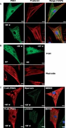
          Hearts from PKN1 knockout (KO) or wild type (WT) littermate control mice were perfused in Langendorff mode and subjected to global ischaemia and reperfusion (I/R). Myocardial infarct size was doubled in PKN1 KO hearts compared to WT hearts. PKN1 was basally phosphorylated on the activation loop Thr 778 PDK1 target site which was unchanged during I/R. However, phosphorylation of p42/p44-MAPK was decreased in KO hearts at baseline and during I/R. In cultured neonatal rat ventricular cardiomyocytes (NRVM) and NRVM transduced with kinase dead (KD) PKN1 K 644R mutant subjected to simulated ischaemia/reperfusion (sI/R), PhosTag ® gel analysis showed net dephosphorylation of PKN1 during sI and early R despite Thr 778 phosphorylation. siRNA knockdown of PKN1 in NRVM significantly decreased cell survival and increased cell injury by sI/R which was reversed by WT- or KD-PKN1 expression. Confocal immunofluorescence analysis of PKN1 in NRVM showed increased localization to the sarcoplasmic reticulum (SR) during sI. GC-MS/MS and immunoblot analysis of PKN1 immunoprecipitates following sI/R confirmed interaction with CamKIIδ. Co-translocation of PKN1 and CamKIIδ to the SR/membrane fraction during sI correlated with phospholamban (PLB) Thr 17 phosphorylation. siRNA knockdown of PKN1 in NRVM resulted in increased basal CamKIIδ activation and increased PLB Thr 17 phosphorylation only during sI. In vivo PLB Thr 17 phosphorylation, Sarco-Endoplasmic Reticulum Ca 2+ ATPase (SERCA2) expression and Junctophilin-2 (Jph2) expression were also basally increased in PKN1 KO hearts. Furthermore, in vivo P-V loop analysis of the beat-to-beat relationship between rate of LV pressure development or relaxation and end diastolic P (EDP) showed mild but significant systolic and diastolic dysfunction with preserved ejection fraction in PKN1 KO hearts.

          Conclusion

          Loss of PKN1 in vivo significantly reduces endogenous cardioprotection and increases myocardial infarct size following I/R injury. Cardioprotection by PKN1 is associated with reduced CamKIIδ-dependent PLB Thr 17 phosphorylation at the SR and therefore may stabilize the coupling of SR Ca 2+ handling and contractile function, independent of its kinase activity.

          Related collections

          Most cited references46

          • Record: found
          • Abstract: found
          • Article: not found

          The extended protein kinase C superfamily.

          Members of the mammalian protein kinase C (PKC) superfamily play key regulatory roles in a multitude of cellular processes, ranging from control of fundamental cell autonomous activities, such as proliferation, to more organismal functions, such as memory. However, understanding of mammalian PKC signalling systems is complicated by the large number of family members. Significant progress has been made through studies based on comparative analysis, which have defined a number of regulatory elements in PKCs which confer specific location and activation signals to each isotype. Further studies on simple organisms have shown that PKC signalling paradigms are conserved through evolution from yeast to humans, underscoring the importance of this family in cellular signalling and giving novel insights into PKC function in complex mammalian systems.
            Bookmark
            • Record: found
            • Abstract: found
            • Article: not found

            CaMKII determines mitochondrial stress responses in heart

            Myocardial cell death is initiated by excessive mitochondrial Ca2+ entry, causing Ca2+ overload, mitochondrial permeability transition pore (mPTP) opening and dissipation of the mitochondrial inner membrane potential (ΔΨm) 1,2 . However, the signaling pathways that control mitochondrial Ca2+ entry through the inner membrane mitochondrial Ca2+ uniporter (MCU) 3–5 are not known. The multifunctional Ca2+ and calmodulin-dependent protein kinase II (CaMKII) is activated in ischemia reperfusion (I/R), myocardial infarction (MI) and neurohumoral injury, common causes of myocardial death and heart failure, suggesting CaMKII could couple disease stress to mitochondrial injury. Here we show that CaMKII promotes mPTP opening and myocardial death by increasing MCU current (IMCU). Mitochondrial-targeted CaMKII inhibitory protein or cyclosporin A (CsA), an mPTP antagonist with clinical efficacy in I/R injury 6 , equivalently prevent mPTP opening, ΔΨm deterioration and diminish mitochondrial disruption and programmed cell death in response to I/R injury. Mice with myocardial and mitochondrial-targeted CaMKII inhibition are resistant to I/R injury, MI and neurohumoral injury, suggesting pathological actions of CaMKII are substantially mediated by increasing IMCU. Our findings identify CaMKII activity as a central mechanism for mitochondrial Ca2+ entry and suggest mitochondrial-targeted CaMKII inhibition could prevent or reduce myocardial death and heart failure dysfunction in response to common experimental forms of pathophysiological stress.
              Bookmark
              • Record: found
              • Abstract: found
              • Article: not found

              Identification of a putative target for Rho as the serine-threonine kinase protein kinase N.

              Rho, a Ras-like small guanosine triphosphatase, has been implicated in cytoskeletal responses to extracellular signals such as lysophosphatidic acid (LPA) to form stress fibers and focal contacts. The form of RhoA bound to guanosine triphosphate directly bound to and activated a serine-threonine kinase, protein kinase N (PKN). Activated RhoA formed a complex with PKN and activated it in COS-7 cells. PKN was phosphorylated in Swiss 3T3 cells stimulated with LPA, and this phosphorylation was blocked by treatment of cells with botulinum C3 exoenzyme. Activation of Rho may be linked directly to a serine-threonine kinase pathway.
                Bookmark

                Author and article information

                Journal
                Cardiovasc Res
                Cardiovasc. Res
                cardiovascres
                Cardiovascular Research
                Oxford University Press
                0008-6363
                1755-3245
                01 January 2018
                16 October 2017
                16 October 2017
                : 114
                : 1
                : 138-157
                Affiliations
                [1 ]Department of Cardiology, School of Cardiovascular Medicine and Sciences, British Heart Foundation Centre for Research Excellence, Faculty of Life Sciences and Medicine, The Rayne Institute, King’s College London, St Thomas’s Hospital, Lambeth Palace Road, London SE1 7EH, UK;
                [2 ]Randall Division of Cell and Molecular Biophysics, King’s College London, New Hunt’s House, Guy’s Hospital Campus, London SE1 1UL, UK;
                [3 ]Division of Cancer Studies, School of Cancer and Pharmaceutical Sciences, Faculty of Life Sciences and Medicine, King’s College London, New Hunt’s House, Guy’s Hospital Campus, London SE1 9RT, UK
                [4 ]Protein Phosphorylation Laboratory, Francis Crick Institute, Lincoln’s Inn Fields, London WC2A 3LY, UK
                Author notes
                Corresponding author. E-mail: richard.heads@ 123456kcl.ac.uk

                Time for primary review: 36 days

                Article
                cvx206
                10.1093/cvr/cvx206
                5815577
                29045568
                82ecc601-5138-406d-88cf-859550635921
                © The Author 2017. Published by Oxford University Press on behalf of the European Society of Cardiology

                This is an Open Access article distributed under the terms of the Creative Commons Attribution License ( http://creativecommons.org/licenses/by/4.0/), which permits unrestricted reuse, distribution, and reproduction in any medium, provided the original work is properly cited.

                History
                : 26 April 2016
                : 17 March 2017
                : 13 October 2017
                Page count
                Pages: 20
                Funding
                Funded by: British Heart Foundation 10.13039/501100000274
                Award ID: #PG/10/045/28276
                Categories
                Original Articles
                Cardiac Biology and Remodelling

                Cardiovascular Medicine
                protein kinase novel 1 (pkn1) • cardioprotection • infarction • camkiiδ • phospholamban

                Comments

                Comment on this article