24
views
0
recommends
+1 Recommend
0 collections
    0
    shares
      • Record: found
      • Abstract: found
      • Article: found
      Is Open Access

      Anogenital Distance Plasticity in Adulthood: Implications for Its Use as a Biomarker of Fetal Androgen Action

      brief-report

      Read this article at

      Bookmark
          There is no author summary for this article yet. Authors can add summaries to their articles on ScienceOpen to make them more accessible to a non-specialist audience.

          Abstract

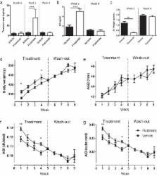
          Androgen action during the fetal masculinization programming window (MPW) determines the maximum potential for growth of androgen-dependent organs (eg, seminal vesicles, prostate, penis, and perineum) and is reflected in anogenital distance (AGD). As such, determining AGD in postnatal life has potential as a lifelong easily accessible biomarker of overall androgen action during the MPW. However, whether the perineum remains androgen responsive in adulthood and thus responds plastically to perturbed androgen drive remains unexplored. To determine this, we treated adult male rats with either the antiandrogen flutamide or the estrogen diethylstilbestrol (DES) for 5 weeks, followed by a 4-week washout period of no treatment. We determined AGD and its correlate anogenital index (AGI) (AGD relative to body weight) at weekly intervals across this period and compared these with normal adult rats (male and female), castrated male rats, and appropriate vehicle controls. These data showed that, in addition to reducing circulating testosterone and seminal vesicle weight, castration significantly reduced AGD (by ∼17%), demonstrating that there is a degree of plasticity in AGD in adulthood. Flutamide treatment increased circulating testosterone yet also reduced seminal vesicle weight due to local antagonism of androgen receptor. Despite this suppression, surprisingly, flutamide treatment had no effect on AGD at any time point. In contrast, although DES treatment suppressed circulating testosterone and reduced seminal vesicle weight, it also induced a significant reduction in AGD (by ∼11%), which returned to normal 1 week after cessation of DES treatment. We conclude that AGD in adult rats exhibits a degree of plasticity, which may be mediated by modulation of local androgen/estrogen action. The implications of these findings regarding the use of AGD as a lifelong clinical biomarker of fetal androgen action are discussed.

          Related collections

          Most cited references23

          • Record: found
          • Abstract: found
          • Article: not found

          Identification in rats of a programming window for reproductive tract masculinization, disruption of which leads to hypospadias and cryptorchidism.

          Becoming a phenotypic male is ultimately determined by androgen-induced masculinization. Disorders of fetal masculinization, resulting in hypospadias or cryptorchidism, are common, but their cause remains unclear. Together with the adult-onset disorders low sperm count and testicular cancer, they can constitute a testicular dysgenesis syndrome (TDS). Although masculinization is well studied, no unifying concept explains normal male reproductive development and its abnormalities, including TDS. We exposed rat fetuses to either anti-androgens or androgens and showed that masculinization of all reproductive tract tissues was programmed by androgen action during a common fetal programming window. This preceded morphological differentiation, when androgen action was, surprisingly, unnecessary. Only within the programming window did blocking androgen action induce hypospadias and cryptorchidism and altered penile length in male rats, all of which correlated with anogenital distance (AGD). Androgen-driven masculinization of females was also confined to the same programming window. This work has identified in rats a common programming window in which androgen action is essential for normal reproductive tract masculinization and has highlighted that measuring AGD in neonatal humans could provide a noninvasive method to predict neonatal and adult reproductive disorders. Based on the timings in rats, we believe the programming window in humans is likely to be 8-14 weeks of gestation.
            Bookmark
            • Record: found
            • Abstract: found
            • Article: not found

            Kiss1-/- mice exhibit more variable hypogonadism than Gpr54-/- mice.

            The G protein-coupled receptor Gpr54 and its ligand metastin (derived from the Kiss1 gene product kisspeptin) are key gatekeepers of sexual maturation. Gpr54 knockout mice demonstrate hypogonadotropic hypogonadism, but until recently, the phenotype of Kiss1 knockout mice was unknown. This report describes the reproductive phenotypes of mice carrying targeted deletions of Kiss1 or Gpr54 on the same genetic background. Both Kiss1 and Gpr54 knockout mice are viable but infertile and have abnormal sexual maturation; the majority of males lack preputial separation, and females have delayed vaginal opening and absence of estrous cycling. Kiss1 and Gpr54 knockout males have significantly smaller testes compared with controls. Gpr54 knockout females have smaller ovaries and uteri than wild-type females. However, Kiss1 knockout females demonstrate two distinct phenotypes: half have markedly reduced gonadal weights similar to those of Gpr54 knockout mice, whereas half exhibit persistent vaginal cornification and have gonadal weights comparable with those of wild-type females. FSH levels in both Kiss1 and Gpr54 knockout males and females are significantly lower than in controls. When injected with mouse metastin 43-52, a Gpr54 agonist, Gpr54 knockout mice fail to increase gonadotropins, whereas Kiss1 knockout mice respond with increased gonadotropin levels. In summary, both Kiss1 and Gpr54 knockout mice have abnormal sexual maturation consistent with hypogonadotropic hypogonadism, although Kiss1 knockout mice appear to be less severely affected than their receptor counterparts. Kiss1 knockout females demonstrate a bimodal phenotypic variability, with some animals having higher gonadal weight, larger vaginal opening, and persistent vaginal cornification.
              Bookmark
              • Record: found
              • Abstract: found
              • Article: not found

              Testicular dysgenesis syndrome: mechanistic insights and potential new downstream effects.

              Reproductive disorders of newborn (cryptorchidism, hypospadias) and young adult males (low sperm counts, testicular germ cell cancer) are common and/or increasing in incidence. It has been hypothesized that these disorders may comprise a testicular dysgenesis syndrome (TDS) with a common origin in fetal life. This has been supported by findings in an animal model of TDS involving fetal exposure to n(dibutyl) phthalate, as well as by new clinical studies. Recent advances in understanding from such studies have led to refinement of the TDS hypothesis, highlighting the central role that deficient androgen production/action during fetal testis development, may play in the origin of downstream disorders.
                Bookmark

                Author and article information

                Journal
                Endocrinology
                Endocrinology
                endo
                endoc
                endo
                Endocrinology
                Endocrine Society (Chevy Chase, MD )
                0013-7227
                1945-7170
                January 2015
                6 November 2014
                6 November 2014
                : 156
                : 1
                : 24-31
                Affiliations
                MRC Centre for Reproductive Health, University of Edinburgh, The Queen's Medical Research Institute, Edinburgh, EH16 4TJ, United Kingdom
                Author notes
                Address all correspondence and requests for reprints to: Professor Lee Smith, MRC Centre for Reproductive Health, University of Edinburgh, The Queen's Medical Research Institute, 47 Little France Crescent, Edinburgh EH16 4TJ, United Kingdom. E-mail: lee.smith@ 123456ed.ac.uk .
                Article
                EN-14-1534
                10.1210/en.2014-1534
                4272396
                25375036
                828e688f-434a-411d-aa6d-77d073e317c7

                This article has been published under the terms of the Creative Commons Attribution License ( CC-BY), which permits unrestricted use, distribution, and reproduction in any medium, provided the original author and source are credited. Copyright for this article is retained by the author(s). Author(s) grant(s) the Endocrine Society the exclusive right to publish the article and identify itself as the original publisher.

                History
                : 27 June 2014
                : 30 October 2014
                Categories
                Brief Reports

                Endocrinology & Diabetes
                Endocrinology & Diabetes

                Comments

                Comment on this article