9
views
0
recommends
+1 Recommend
0 collections
    0
    shares
      • Record: found
      • Abstract: found
      • Article: found
      Is Open Access

      Ablation of the canonical testosterone production pathway via knockout of the steroidogenic enzyme HSD17B3, reveals a novel mechanism of testicular testosterone production

      research-article

      Read this article at

      Bookmark
          There is no author summary for this article yet. Authors can add summaries to their articles on ScienceOpen to make them more accessible to a non-specialist audience.

          Abstract

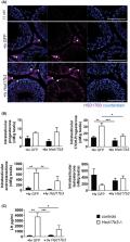
          Male development, fertility, and lifelong health are all androgen‐dependent. Approximately 95% of circulating testosterone is synthesized by the testis and the final step in this canonical pathway is controlled by the activity of the hydroxysteroid‐dehydrogenase‐17‐beta‐3 (HSD17B3). To determine the role of HSD17B3 in testosterone production and androgenization during male development and function we have characterized a mouse model lacking HSD17B3. The data reveal that developmental masculinization and fertility are normal in mutant males. Ablation of HSD17B3 inhibits hyperstimulation of testosterone production by hCG, although basal testosterone levels are maintained despite the absence of HSD17B3. Reintroduction of HSD17B3 via gene‐delivery to Sertoli cells in adulthood partially rescues the adult phenotype, showing that, as in development, different cell‐types in the testis are able to work together to produce testosterone. Together, these data show that HS17B3 acts as a rate‐limiting‐step for the maximum level of testosterone production by the testis but does not control basal testosterone production. Measurement of other enzymes able to convert androstenedione to testosterone identifies HSD17B12 as a candidate enzyme capable of driving basal testosterone production in the testis. Together, these findings expand our understanding of testosterone production in males.

          Related collections

          Most cited references45

          • Record: found
          • Abstract: found
          • Article: not found

          Identification in rats of a programming window for reproductive tract masculinization, disruption of which leads to hypospadias and cryptorchidism.

          Becoming a phenotypic male is ultimately determined by androgen-induced masculinization. Disorders of fetal masculinization, resulting in hypospadias or cryptorchidism, are common, but their cause remains unclear. Together with the adult-onset disorders low sperm count and testicular cancer, they can constitute a testicular dysgenesis syndrome (TDS). Although masculinization is well studied, no unifying concept explains normal male reproductive development and its abnormalities, including TDS. We exposed rat fetuses to either anti-androgens or androgens and showed that masculinization of all reproductive tract tissues was programmed by androgen action during a common fetal programming window. This preceded morphological differentiation, when androgen action was, surprisingly, unnecessary. Only within the programming window did blocking androgen action induce hypospadias and cryptorchidism and altered penile length in male rats, all of which correlated with anogenital distance (AGD). Androgen-driven masculinization of females was also confined to the same programming window. This work has identified in rats a common programming window in which androgen action is essential for normal reproductive tract masculinization and has highlighted that measuring AGD in neonatal humans could provide a noninvasive method to predict neonatal and adult reproductive disorders. Based on the timings in rats, we believe the programming window in humans is likely to be 8-14 weeks of gestation.
            Bookmark
            • Record: found
            • Abstract: found
            • Article: not found

            Characteristics of secondary, primary, and compensated hypogonadism in aging men: evidence from the European Male Ageing Study.

            The diagnosis of late-onset hypogonadism (LOH) in older men with age-related declines in testosterone (T) is currently not well characterized. Our objective was to investigate whether different forms of hypogonadism can be distinguished among aging men. The study was a cross-sectional survey on 3369 community-dwelling men aged 40-79 yr in eight European centers. Four groups of subjects were defined: eugonadal (normal T and normal LH), secondary (low T and low/normal LH), primary (low T and elevated LH), and compensated (normal T and elevated LH) hypogonadism. Relationships between the defined gonadal status with potential risk factors and clinical symptoms were investigated by multilevel regression models. Among the men, 11.8, 2.0, and 9.5% were classified into the secondary, primary, and compensated hypogonadism categories, respectively. Older men were more likely to have primary [relative risk ratio (RRR) = 3.04; P < 0.001] and compensated (RRR = 2.41; P < 0.001) hypogonadism. Body mass index of 30 kg/m(2) or higher was associated with secondary hypogonadism (RRR = 8.74; P < 0.001). Comorbidity was associated with both secondary and primary hypogonadism. Sexual symptoms were more prevalent in secondary and primary hypogonadism, whereas physical symptoms were more likely in compensated hypogonadism. Symptomatic elderly men considered to have LOH can be differentiated on the basis of endocrine and clinical features and predisposing risk factors. Secondary hypogonadism is associated with obesity and primary hypogonadism predominately with age. Compensated hypogonadism can be considered a distinct clinical state associated with aging. Classification of LOH into different categories by combining LH with T may improve the diagnosis and management of LOH.
              Bookmark
              • Record: found
              • Abstract: found
              • Article: not found

              Differential regulation of KiSS-1 mRNA expression by sex steroids in the brain of the male mouse.

              Kisspeptins are products of the Kiss1 gene, which bind to GPR54, a G protein-coupled receptor. Kisspeptins and GPR54 have been implicated in the neuroendocrine regulation of GnRH secretion. To test the hypothesis that testosterone regulates Kiss1 gene expression, we compared the expression of KiSS-1 mRNA among groups of intact, castrated, and castrated/testosterone (T)-treated male mice. In the arcuate nucleus (Arc), castration resulted in a significant increase in KiSS-1 mRNA, which was completely reversed with T replacement, whereas in the anteroventral periventricular nucleus, the results were the opposite, i.e. castration decreased and T increased KiSS-1 mRNA expression. In the Arc, the effects of T on KiSS-1 mRNA were completely mimicked by estrogen but only partially mimicked by dihydrotestosterone, a nonaromatizable androgen, suggesting that both estrogen receptor (ER) and androgen receptor (AR) play a role in T-mediated regulation of KiSS-1. Studies of the effects of T on KiSS-1 expression in mice with either a deletion of the ERalpha or a hypomorphic allele to the AR revealed that the effects of T are mediated by both ERalpha and AR pathways, which was confirmed by the presence of either ERalpha or AR coexpression in most KiSS-1 neurons in the Arc. These observations suggest that KiSS-1 neurons in the Arc, whose transcriptional activity is inhibited by T, are targets for the negative feedback regulation of GnRH secretion, whereas KiSS-1 neurons in the anteroventral periventricular nucleus, whose activity is stimulated by T, may mediate other T-dependent processes.
                Bookmark

                Author and article information

                Contributors
                L.B.Smith@newcastle.edu.au
                Journal
                FASEB J
                FASEB J
                10.1096/(ISSN)1530-6860
                FSB2
                The FASEB Journal
                John Wiley and Sons Inc. (Hoboken )
                0892-6638
                1530-6860
                18 June 2020
                August 2020
                : 34
                : 8 ( doiID: 10.1096/fsb2.v34.8 )
                : 10373-10386
                Affiliations
                [ 1 ] School of Environmental and Life Sciences University of Newcastle Callaghan NSW Australia
                [ 2 ] MRC Centre for Reproductive Health University of Edinburgh The Queen’s Medical Research Institute Edinburgh UK
                [ 3 ] Department of Growth and Reproduction, Rigshospitalet University of Copenhagen Copenhagen Denmark
                [ 4 ] International Centre for Research and Research Training in Endocrine Disruption of Male Reproduction and Child Health, Rigshospitalet University of Copenhagen Copenhagen Denmark
                [ 5 ] Institute of Biodiversity, Animal Health, and Comparative Medicine University of Glasgow Glasgow UK
                [ 6 ] Department of Genetic Medicine and Development Faculty of Medicine University of Geneva Geneva Switzerland
                Author notes
                [*] [* ] Correspondence

                Lee B. Smith, ,School of Environmental and Life Sciences, University of Newcastle, Callaghan, NSW 2308, Australia.

                Email: L.B.Smith@ 123456newcastle.edu.au

                Article
                FSB220705
                10.1096/fj.202000361R
                7496839
                32557858
                50264314-8bee-48ce-8119-3cd86d548ab0
                © 2020 The Authors. The FASEB Journal published by Wiley Periodicals LLC on behalf of Federation of American Societies for Experimental Biology

                This is an open access article under the terms of the http://creativecommons.org/licenses/by/4.0/ License, which permits use, distribution and reproduction in any medium, provided the original work is properly cited.

                History
                : 24 February 2020
                : 07 May 2020
                : 20 May 2020
                Page count
                Figures: 6, Tables: 2, Pages: 14, Words: 16939
                Funding
                Funded by: Department of Health | National Health and Medical Research Council (NHMRC) , open-funder-registry 10.13039/501100000925;
                Award ID: APP1158344
                Funded by: RCUK | Medical Research Council (MRC) , open-funder-registry 10.13039/501100000265;
                Award ID: MR/N002970/1
                Categories
                Research Article
                Research Articles
                Custom metadata
                2.0
                August 2020
                Converter:WILEY_ML3GV2_TO_JATSPMC version:5.9.0 mode:remove_FC converted:11.09.2020

                Molecular biology
                androgens,hsd17b3,hsd17b12,leydig cell,testis,testosterone
                Molecular biology
                androgens, hsd17b3, hsd17b12, leydig cell, testis, testosterone

                Comments

                Comment on this article