5
views
0
recommends
+1 Recommend
0 collections
    0
    shares
      • Record: found
      • Abstract: found
      • Article: found
      Is Open Access

      Extra-mitochondrial mouse frataxin and its implications for mouse models of Friedreich’s ataxia

      research-article

      Read this article at

      Bookmark
          There is no author summary for this article yet. Authors can add summaries to their articles on ScienceOpen to make them more accessible to a non-specialist audience.

          Abstract

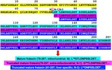
          Mature frataxin is essential for the assembly of iron–sulfur cluster proteins including a number of mitochondrial enzymes. Reduced levels of mature frataxin (81-20) in human subjects caused by the genetic disease Friedreich’s ataxia results in decreased mitochondrial function, neurodegeneration, and cardiomyopathy. Numerous studies of mitochondrial dysfunction have been conducted using mouse models of frataxin deficiency. However, mouse frataxin that is reduced in these models, is assumed to be mature frataxin (78-207) by analogy with human mature frataxin (81-210). Using immunoaffinity purification coupled with liquid chromatography-high resolution tandem mass spectrometry, we have discovered that mature frataxin in mouse heart (77%), brain (86%), and liver (47%) is predominantly a 129-amino acid truncated mature frataxin (79-207) in which the N-terminal lysine residue has been lost. Mature mouse frataxin (78-207) only contributes 7–15% to the total frataxin protein present in mouse tissues. We have also found that truncated mature frataxin (79-207) is present primarily in the cytosol of mouse liver; whereas, frataxin (78-207) is primarily present in the mitochondria. These findings, which provide support for the role of extra-mitochondrial frataxin in the etiology of Friedreich’s ataxia, also have important implications for studies of mitochondrial dysfunction conducted in mouse models of frataxin deficiency.

          Related collections

          Most cited references35

          • Record: found
          • Abstract: found
          • Article: not found

          Global analysis of the mitochondrial N-proteome identifies a processing peptidase critical for protein stability.

          Many mitochondrial proteins are synthesized with N-terminal presequences that are removed by specific peptidases. The N-termini of the mature proteins and thus peptidase cleavage sites have only been determined for a small fraction of mitochondrial proteins and yielded a controversial situation for the cleavage site specificity of the major mitochondrial processing peptidase (MPP). We report a global analysis of the N-proteome of yeast mitochondria, revealing the N-termini of 615 different proteins. Significantly more proteins than predicted contained cleavable presequences. We identified the intermediate cleaving peptidase Icp55, which removes an amino acid from a characteristic set of MPP-generated N-termini, solving the controversial situation of MPP specificity and suggesting that Icp55 converts instable intermediates into stable proteins. Our results suggest that Icp55 is critical for stabilization of the mitochondrial proteome and illustrate how the N-proteome can serve as rich source for a systematic analysis of mitochondrial protein targeting, cleavage and turnover.
            Bookmark
            • Record: found
            • Abstract: found
            • Article: not found

            The role of mitochondria in cellular iron-sulfur protein biogenesis and iron metabolism.

            Mitochondria play a key role in iron metabolism in that they synthesize heme, assemble iron-sulfur (Fe/S) proteins, and participate in cellular iron regulation. Here, we review the latter two topics and their intimate connection. The mitochondrial Fe/S cluster (ISC) assembly machinery consists of 17 proteins that operate in three major steps of the maturation process. First, the cysteine desulfurase complex Nfs1-Isd11 as the sulfur donor cooperates with ferredoxin-ferredoxin reductase acting as an electron transfer chain, and frataxin to synthesize an [2Fe-2S] cluster on the scaffold protein Isu1. Second, the cluster is released from Isu1 and transferred toward apoproteins with the help of a dedicated Hsp70 chaperone system and the glutaredoxin Grx5. Finally, various specialized ISC components assist in the generation of [4Fe-4S] clusters and cluster insertion into specific target apoproteins. Functional defects of the core ISC assembly machinery are signaled to cytosolic or nuclear iron regulatory systems resulting in increased cellular iron acquisition and mitochondrial iron accumulation. In fungi, regulation is achieved by iron-responsive transcription factors controlling the expression of genes involved in iron uptake and intracellular distribution. They are assisted by cytosolic multidomain glutaredoxins which use a bound Fe/S cluster as iron sensor and additionally perform an essential role in intracellular iron delivery to target metalloproteins. In mammalian cells, the iron regulatory proteins IRP1, an Fe/S protein, and IRP2 act in a post-transcriptional fashion to adjust the cellular needs for iron. Thus, Fe/S protein biogenesis and cellular iron metabolism are tightly linked to coordinate iron supply and utilization. This article is part of a Special Issue entitled: Cell Biology of Metals. Copyright © 2012 Elsevier B.V. All rights reserved.
              Bookmark
              • Record: found
              • Abstract: found
              • Article: not found

              Prevention and reversal of severe mitochondrial cardiomyopathy by gene therapy in a mouse model of Friedreich's ataxia.

              Cardiac failure is the most common cause of mortality in Friedreich's ataxia (FRDA), a mitochondrial disease characterized by neurodegeneration, hypertrophic cardiomyopathy and diabetes. FRDA is caused by reduced levels of frataxin (FXN), an essential mitochondrial protein involved in the biosynthesis of iron-sulfur (Fe-S) clusters. Impaired mitochondrial oxidative phosphorylation, bioenergetics imbalance, deficit of Fe-S cluster enzymes and mitochondrial iron overload occur in the myocardium of individuals with FRDA. No treatment exists as yet for FRDA cardiomyopathy. A conditional mouse model with complete frataxin deletion in cardiac and skeletal muscle (Mck-Cre-Fxn(L3/L-) mice) recapitulates most features of FRDA cardiomyopathy, albeit with a more rapid and severe course. Here we show that adeno-associated virus rh10 vector expressing human FXN injected intravenously in these mice fully prevented the onset of cardiac disease. Moreover, later administration of the frataxin-expressing vector, after the onset of heart failure, was able to completely reverse the cardiomyopathy of these mice at the functional, cellular and molecular levels within a few days. Our results demonstrate that cardiomyocytes with severe energy failure and ultrastructure disorganization can be rapidly rescued and remodeled by gene therapy and establish the preclinical proof of concept for the potential of gene therapy in treating FRDA cardiomyopathy.
                Bookmark

                Author and article information

                Contributors
                ianblair@upenn.edu
                Journal
                Sci Rep
                Sci Rep
                Scientific Reports
                Nature Publishing Group UK (London )
                2045-2322
                25 September 2020
                25 September 2020
                2020
                : 10
                : 15788
                Affiliations
                [1 ]GRID grid.25879.31, ISNI 0000 0004 1936 8972, Penn Medicine/CHOP Center of Excellence in Friedreich’s Ataxia, , University of Pennsylvania, ; Philadelphia, PA 19104 USA
                [2 ]GRID grid.25879.31, ISNI 0000 0004 1936 8972, Center of Excellence in Environmental Toxicology, Department of Systems Pharmacology and Translational Therapeutics, Perelman School of Medicine, , University of Pennsylvania, ; Philadelphia, PA 19104 USA
                [3 ]GRID grid.25879.31, ISNI 0000 0004 1936 8972, Departments of Pediatrics and Neurology, Perelman School of Medicine, , University of Pennsylvania, ; Philadelphia, PA 19104 USA
                Author information
                http://orcid.org/0000-0003-0366-8658
                Article
                72884
                10.1038/s41598-020-72884-w
                7519113
                32978498
                8068836e-bee4-4b52-9ce7-37f17dfbdaf7
                © The Author(s) 2020

                Open Access This article is licensed under a Creative Commons Attribution 4.0 International License, which permits use, sharing, adaptation, distribution and reproduction in any medium or format, as long as you give appropriate credit to the original author(s) and the source, provide a link to the Creative Commons licence, and indicate if changes were made. The images or other third party material in this article are included in the article's Creative Commons licence, unless indicated otherwise in a credit line to the material. If material is not included in the article's Creative Commons licence and your intended use is not permitted by statutory regulation or exceeds the permitted use, you will need to obtain permission directly from the copyright holder. To view a copy of this licence, visit http://creativecommons.org/licenses/by/4.0/.

                History
                : 13 January 2020
                : 28 August 2020
                Funding
                Funded by: National Institutes of Health
                Award ID: T32ES019851
                Award ID: P30ES013508
                Award ID: R01FD006029
                Award Recipient :
                Categories
                Article
                Custom metadata
                © The Author(s) 2020

                Uncategorized
                isoenzymes,multienzyme complexes,neurodegenerative diseases
                Uncategorized
                isoenzymes, multienzyme complexes, neurodegenerative diseases

                Comments

                Comment on this article