69
views
0
recommends
+1 Recommend
1 collections
    0
    shares

      To submit to this journal, click here

      • Record: found
      • Abstract: found
      • Article: found
      Is Open Access

      Méningiome intracrânien multiple: expérience du service de neurochirurgie CHU Avicenne Rabat - Salé, à propos de 4 cas et revue de la literature Translated title: Multiple intracranial meningioma: experience of the neurosurgery serice of Avicenna Hospital Rabat - Salé, about 4 cases and review of the literature

      case-report

      Read this article at

      ScienceOpenPublisherPMC
      Bookmark
          There is no author summary for this article yet. Authors can add summaries to their articles on ScienceOpen to make them more accessible to a non-specialist audience.

          Abstract

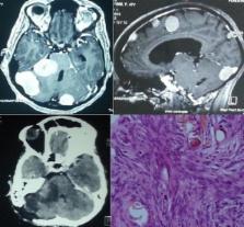
          Les méningiomes intracrâniennes multiples sont définies comme la présence d'au moins deux méningiomes sur des sites intracrâniens différents et ceci en absence de neurofibromatose. C'est une tumeur rare dont la prévalence varie entre 1-10%. Le but de notre travail était de décrire les caractéristiques cliniques, radiologiques, histologiques d'une série de 4 patients porteurs de méningiome multiple et en déduire les facteurs de risques de survenue de cette pathologie. Préciser la qualité d'exérèse chirurgicale de la lésion selon la classification de Simpson. Rapporter les suites postopératoires ainsi que le suivie à long termes des patients afin de préciser leur qualité de vie. Il s'agit d'une étude rétrospective portant sur 4 cas de Méningiomes intracrâniens multiples sur 174 patients opérés pour méningiome au CHU Avicenne entre Janvier 2000 à Décembre 2013. En s'aidant des données cliniques, imageries, chirurgicales, histologiques mentionnée dans le dossier médical de chaque patient. Notre série est constitué de 4 patients (3 femmes pour 1 homme), d'un âge allant de 42-50 ans (moyenne d’âge= 45,5 ans). Nous avons identifié 21 méningiomes (17 en sus tentoriel et 4 en sous tentoriel), aucun cas de décès ni d'infection postopératoire dans notre échantillon. Le pronostic reste bon malgré le nombre de lésion nécessitant parfois plusieurs interventions chirurgicales.

          Most cited references18

          • Record: found
          • Abstract: found
          • Article: not found

          Radiation-induced meningiomas: experience at the Mount Sinai Hospital and review of the literature.

          From the records of The Mount Sinai Hospital, seven cases which met established criteria for radiation-induced meningiomas were identified. This represents the largest series of radiogenic meningiomas documented in North America and includes both intracranial and intraspinal tumors. The records and pathological specimens were reviewed and these data analyzed with other cases retrieved from the world literature. This study reveals that radiation-induced meningiomas can be categorized into three groups based on the amount of radiation administered: 1) low dose; 2) moderate dose and miscellaneous; and 3) high dose. The overwhelming majority of cases had received low-dose irradiation (800 rad) to the scalp for tinea capitis and the second largest group resulted from high-dose irradiation for primary brain tumors (greater than 2000 rad). The unique features distinguishing radiation-induced meningiomas from other meningiomas are reviewed. Although histologically atypical tumors were common in this series, overt malignancy was not encountered. The preoperative management of these lesions should include angiography to evaluate for large-vessel occlusive vasculopathy, a known association of meningiomas induced by high-dose irradiation. Given the propensity these tumors possess for recurrence, a wide bony and dural margin is recommended at surgical resection.
            Bookmark
            • Record: found
            • Abstract: found
            • Article: found
            Is Open Access

            Natural history of multiple meningiomas

            Background: Asymptomatic solitary meningiomas are typically managed with clinical and radiographic follow-up. Multiple meningiomas represents a clinical entity distinct from solitary meningiomas and can be sporadic, radiation-induced, associated with neurofibromatosis, or exhibit other familial inheritance. The growth rate for multiple meningiomas is not known and therefore management of these complicated patients can be difficult. Methods: A retrospective chart review was performed on 12 patients with a total of 55 meningiomas. Patients with neurofibromatosis were not included. Serial enhanced magnetic resonance imaging was used to determine tumor growth rates. Treatment history was also reviewed and included for analysis. Results: Analysis of all 55 tumors demonstrated an average rate of growth of 0.46 cm3/year (range: −0.57-2.94 cm3/year). In the 23 tumors that received no treatment, the average rate of growth was 0.34 cm3/year (range: −0.03-1.8 cm3/year). Ten of the 23 tumors that received no treatment had no history of cranial irradiation. This group demonstrated a growth rate of 0.44 cm3/year (range: −0.01-1.8 cm3/year). Linear regression analysis did not yield any significant relationship between tumor burden and rates of growth. Conclusion: Tumor growth rates in patients with multiple meningiomas did not appear to be higher than reported rates for incidentally found solitary meningiomas. As such, asymptomatic multiple meningioma patients should be managed with clinical and radiographic follow-up.
              Bookmark
              • Record: found
              • Abstract: found
              • Article: not found

              Estrogen and progesterone receptors in meningiomas in relation to clinical and pathologic features.

              Tumor estradiol and progesterone binding sites were studied in 34 patients with meningioma. Twenty of the meningiomas contained very low titers (mean, 45 fmol/g of tumor; range, 0-201 fmol/g of tumor) of a nonspecific cytoplasmic [3H]estradiol binding component, whereas 26 of the tumors contained high titers of specific high-affinity cytosol [3H]promegestone (R5020; progesterone) binding sites (mean, 1476 fmol/g of tumor; range, 0-8328 fmol/g of tumor). No nuclear binding activity for [3H]estradiol could be detected in 12 of the 34 meningiomas studied, irrespective of the progesterone binding activity. There was no correlation between high progesterone binding activity and the age or the sex of the patient, nor between tumor location and cellular mitotic index. However, progesterone binding activity was present more frequently in meningothelial (95%, 18/21 patients) than in transitional (55%, 5/9 patients) or fibroplastic (25%, 1/4 patients) tumor histologic types. These data suggest that the cellular biosynthesis of the progesterone binding component in meningiomas is not estrogen regulated as it is in other classic estrogen target tissues, such as the breast.
                Bookmark

                Author and article information

                Journal
                Pan Afr Med J
                Pan Afr Med J
                PAMJ
                The Pan African Medical Journal
                The African Field Epidemiology Network
                1937-8688
                06 July 2014
                2014
                : 18
                : 204
                Affiliations
                [1 ]Service de neurochirurgie CHU Avicenne, Rabat-Salé, Université Mohammed V Souissi, Rabat, Maroc
                Author notes
                [& ]Corresponding author: Ben Ousmanou Djoubairou, Service de neurochirurgie CHU Avicenne, Rabat-Salé, Université Mohammed V Souissi, Rabat, Maroc
                Article
                PAMJ-18-204
                10.11604/pamj.2014.18.204.4811
                4237563
                8052835e-dd27-4993-8b1c-830c464cf05e
                © Ben Ousmanou Djoubairou et al.

                The Pan African Medical Journal - ISSN 1937-8688. This is an Open Access article distributed under the terms of the Creative Commons Attribution License which permits unrestricted use, distribution, and reproduction in any medium, provided the original work is properly cited.

                History
                : 14 June 2014
                : 30 June 2014
                Categories
                Case Series

                Medicine
                méningiome multiple,exérèse chirurgicale,neurofibromatose,multiple meningioma,surgical excision,neurofibromatosis

                Comments

                Comment on this article