31
views
0
recommends
+1 Recommend
0 collections
    0
    shares
      • Record: found
      • Abstract: found
      • Article: found
      Is Open Access

      In vivo Imaging of Intact Drosophila Larvae at Sub-cellular Resolution

      research-article

      Read this article at

      Bookmark
          There is no author summary for this article yet. Authors can add summaries to their articles on ScienceOpen to make them more accessible to a non-specialist audience.

          Abstract

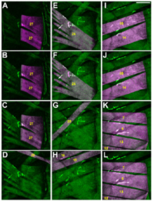
          Recent improvements in optical imaging, genetically encoded fluorophores and genetic tools allowing efficient establishment of desired transgenic animal lines have enabled biological processes to be studied in the context of a living, and in some instances even behaving, organism. In this protocol we will describe how to anesthetize intact Drosophila larvae, using the volatile anesthetic desflurane, to follow the development and plasticity of synaptic populations at sub-cellular resolution 1-3. While other useful methods to anesthetize Drosophila melanogaster larvae have been previously described 4,5,6,7,8, the protocol presented herein demonstrates significant improvements due to the following combined key features: (1) A very high degree of anesthetization; even the heart beat is arrested allowing for lateral resolution of up to 150 nm 1, (2) a high survival rate of > 90% per anesthetization cycle, permitting the recording of more than five time-points over a period of hours to days 2 and (3) a high sensitivity enabling us in 2 instances to study the dynamics of proteins expressed at physiological levels. In detail, we were able to visualize the postsynaptic glutamate receptor subunit GluR-IIA expressed via the endogenous promoter 1 in stable transgenic lines and the exon trap line FasII-GFP 1. (4) In contrast to other methods 4,7 the larvae can be imaged not only alive, but also intact (i.e. non-dissected) allowing observation to occur over a number of days 1. The accompanying video details the function of individual parts of the in vivo imaging chamber 2,3, the correct mounting of the larvae, the anesthetization procedure, how to re-identify specific positions within a larva and the safe removal of the larvae from the imaging chamber.

          Related collections

          Most cited references8

          • Record: found
          • Abstract: found
          • Article: not found

          Glutamate receptor dynamics organizing synapse formation in vivo.

          Insight into how glutamatergic synapses form in vivo is important for understanding developmental and experience-triggered changes of excitatory circuits. Here, we imaged postsynaptic densities (PSDs) expressing a functional, GFP-tagged glutamate receptor subunit (GluR-IIA(GFP)) at neuromuscular junctions of Drosophila melanogaster larvae for several days in vivo. New PSDs, associated with functional and structural presynaptic markers, formed independently of existing synapses and grew continuously until reaching a stable size within hours. Both in vivo photoactivation and photobleaching experiments showed that extrasynaptic receptors derived from diffuse, cell-wide pools preferentially entered growing PSDs. After entering PSDs, receptors were largely immobilized. In comparison, other postsynaptic proteins tested (PSD-95, NCAM and PAK homologs) exchanged faster and with no apparent preference for growing synapses. We show here that new glutamatergic synapses form de novo and not by partitioning processes from existing synapses, suggesting that the site-specific entry of particular glutamate receptor complexes directly controls the assembly of individual PSDs.
            Bookmark
            • Record: found
            • Abstract: found
            • Article: not found

            Watching a synapse grow: noninvasive confocal imaging of synaptic growth in Drosophila.

            The glutamatergic neuromuscular junction (NMJ) in Drosophila adds new boutons and branches during larval development. We generated transgenic fruit flies that express a novel green fluorescent membrane protein at the postsynaptic specialization, allowing for repeated noninvasive confocal imaging of synapses in live, developing larvae. As synapses grow, existing synaptic boutons stretch apart and new boutons insert between them; in addition, new boutons are added at the ends of existing strings of boutons. Some boutons are added de novo, while others bud from existing boutons. New branches form as multiple boutons bud from existing boutons. Nascent boutons contain active zones, T bars, and synaptic vesicles; we observe no specialized growth structures. Some new boutons exhibit a lower level of Fasciclin II, suggesting that the levels of this synaptic cell adhesion molecule vary locally during synaptic growth.
              Bookmark
              • Record: found
              • Abstract: found
              • Article: not found

              Imaging axonal transport of mitochondria.

              Neuronal mitochondria need to be transported and distributed in axons and dendrites in order to ensure an adequate energy supply and provide sufficient Ca(2+) buffering in each portion of these highly extended cells. Errors in mitochondrial transport are implicated in neurodegenerative diseases. Here we present useful tools to analyze axonal transport of mitochondria both in vitro in cultured rat neurons and in vivo in Drosophila larval neurons. These methods enable investigators to take advantage of both systems to study the properties of mitochondrial motility under normal or pathological conditions.
                Bookmark

                Author and article information

                Journal
                J Vis Exp
                J Vis Exp
                JoVE
                Journal of Visualized Experiments : JoVE
                MyJove Corporation
                1940-087X
                2010
                10 September 2010
                10 September 2010
                : 43
                : 2249
                Affiliations
                Junior Research Group Synaptic Plasticity, Hertie Institute for Clinical Brain Research, University of Tübingen
                Graduate School of Cellular and Molecular Neuroscience, University of Tübingen
                Author notes

                Correspondence to: Tobias M. Rasse at tobias.rasse@ 123456uni-tuebingen.de

                Article
                2249
                10.3791/2249
                3157860
                20864927
                7f96f5e1-1d8e-4652-a037-548f6d994a23
                Copyright © 2010, Journal of Visualized Experiments

                This is an open-access article distributed under the terms of the Creative Commons Attribution License, which permits unrestricted use, distribution, and reproduction in any medium, provided the original work is properly cited.

                History
                Categories
                Basic Protocols

                Uncategorized
                Uncategorized

                Comments

                Comment on this article