26
views
0
recommends
+1 Recommend
0 collections
    0
    shares
      • Record: found
      • Abstract: found
      • Article: found
      Is Open Access

      Morphometric analysis of small arteries in the human retina using adaptive optics imaging: relationship with blood pressure and focal vascular changes

      research-article

      Read this article at

      Bookmark
          There is no author summary for this article yet. Authors can add summaries to their articles on ScienceOpen to make them more accessible to a non-specialist audience.

          Abstract

          Objectives:

          The wall-to-lumen ratio (WLR) of retinal arteries is a recognized surrogate of end-organ damage due to aging and/or arterial hypertension. However, parietal morphometry remains difficult to assess in vivo. Recently, it was shown that adaptive optics retinal imaging can resolve parietal structures of retinal arterioles in humans in vivo. Here, using adaptive optics retinal imaging, we investigated the variations of parietal thickness of small retinal arteries with blood pressure and focal vascular damage.

          Methods:

          Adaptive optics imaging of the superotemporal retinal artery was done in 49 treatment-naive individuals [mean age (±SD) 44.9 years (±14); mean systolic pressure 132 mmHg (±22)]. Semi-automated segmentation allowed extracting parietal thickness and lumen diameter. In a distinct cohort, adaptive optics images of arteriovenous nicking (AVN; n = 12) and focal arteriolar narrowing (FAN; n = 10) were also analyzed qualitatively and quantitatively.

          Results:

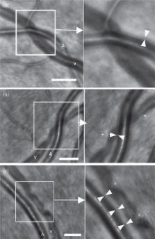
          In the cohort of treatment-naive individuals, by multiple regression taking into account age, body mass index, mean, systolic, diastolic and pulse blood pressure, the WLR was found positively correlated to mean blood pressure and age which in combination accounted for 43% of the variability of WLR. In the cohort of patients with focal vascular damage, neither FANs or AVNs showed evidence of parietal growth; instead, at sites of FANs, decreased outer diameter suggestive of vasoconstriction was consistently found, while at sites of AVNs venous narrowing could be seen in the absence of arteriovenous contact.

          Conclusion:

          High resolution imaging of retinal vessels by adaptive optics allows quantitative microvascular phenotyping, which may contribute to a better understanding and management of hypertensive retinopathy.

          Related collections

          Most cited references23

          • Record: found
          • Abstract: found
          • Article: not found

          Retinal vessel diameters and risk of hypertension: the Rotterdam Study.

          Generalized retinal arteriolar narrowing is an important sign of systemic hypertension, and a lower arteriolar:venular diameter ratio predicts the risk of hypertension. We investigated whether this association was based on arteriolar or venular diameters or both. This study was based on the prospective population-based Rotterdam Study (1990-1993) and included 1900 participants (> or =55 years of age) of whom 739 persons had normal blood pressure (systolic or =140 mm Hg or diastolic blood pressure > or =90 mm Hg or use of antihypertensive medication. Adjusted for age, gender, follow-up time, body mass index, smoking, diabetes mellitus, total and high-density lipoprotein cholesterol, C-reactive protein, and intima-media thickness, arteriolar narrowing was associated with an increased risk of hypertension (odds ratio per SD: 1.38; 95% CI, 1.23 to 1.55); for venular narrowing this was less striking (OR: 1.17; 95% CI, 1.04 to 1.32). Each SD decrease in the arteriolar:venular diameter ratio significantly increased the risk of hypertension by 24%. To examine the effect of baseline blood pressure, we stratified persons into those with "normal blood pressure" or "prehypertension." Within these strata, arteriolar narrowing was still related to incident hypertension. These data show that both retinal arteriolar and venular narrowing may precede the development of systemic hypertension.
            Bookmark
            • Record: found
            • Abstract: found
            • Article: not found

            Retinal microvascular abnormalities and risk of lacunar stroke: Atherosclerosis Risk in Communities Study.

            The retinal microvasculature reflects cumulative small-vessel damage from hypertension and other vascular processes. No study has prospectively examined retinal findings in relation to the incidence of clinical lacunar stroke in comparison with other ischemic stroke subtypes. In 10 496 adults initially free of stroke, we related retinal findings imaged during 1993 to 1995 with the incidence of hospitalized ischemic strokes through 2005. During a median of 11.2 years of follow-up, 338 incident ischemic strokes occurred (66 lacunar, 192 nonlacunar thrombotic, and 80 cardioembolic). Generalized arteriolar narrowing as measured by the central retinal arteriole equivalent was associated with an increased incidence of lacunar stroke (multivariate-adjusted hazard ratio [HR] per 1-SD decrement of central retinal arteriole equivalent=1.67; 95% CI, 1.23-2.26) but was not associated with other ischemic stroke subtypes. Generalized venular widening as measured by the central retinal venule equivalent was also positively associated with only lacunar stroke (multivariate-adjusted HR per 1-SD increment=1.44; 95% CI, 1.09-1.91). Retinal microvascular abnormalities were positively associated with lacunar stroke incidence (HR for focal arteriolar narrowing=2.22; 95% CI, 1.11=4.48; for arteriovenous nicking, HR=2.38; 95% CI, 1.20-4.71), whereas retinopathy signs (microaneurysms, retinal hemorrhages, and others) were positively associated with nonlacunar thrombotic (HR=2.41; 95% CI, 1.47-3.95) and cardioembolic (HR=2.25; 95% CI, 1.09-4.65) stroke incidence. A narrower central retinal arteriole equivalent, wider central retinal venule equivalent, focal arteriolar narrowing, and arteriovenous nicking were predictive of lacunar stroke. Retinal imaging is useful in understanding the pathophysiology and mechanisms of cerebral small-vessel disease.
              Bookmark
              • Record: found
              • Abstract: not found
              • Article: not found

              Small artery structure in hypertension. Dual processes of remodeling and growth.

                Bookmark

                Author and article information

                Journal
                J Hypertens
                J. Hypertens
                JHYPE
                Journal of Hypertension
                Lippincott Williams & Wilkins
                0263-6352
                1473-5598
                April 2014
                10 March 2014
                : 32
                : 4
                : 890-898
                Affiliations
                [a ]Clinical Investigation Center 503, Centre Hospitalier National des Quinze-Vingts, INSERM and Université Pierre et Marie Curie-Paris6
                [b ]Preventive Cardiovascular Unit, Institute of Cardiometabolism and Nutrition, Pitié-Salpêtrière Hospital, Assistance Publique-Hôpitaux de Paris
                [c ]Université Paris-Sud, Hôpital Bicêtre, Assistance Publique-Hôpitaux de Paris,
                [d ]Institut Supérieur d’Electronique de Paris, Paris, France
                Author notes
                Correspondence to Michel Paques, MD, PhD, Clinical Investigation Center 503, Centre Hospitalier National des Quinze-Vingts, 28 rue de Charenton 75012 Paris, France. Tel: +33 1 40 02 14 15; fax: +33 1 40 02 14 99; e-mail: michel.paques@ 123456gmail.com
                Article
                10.1097/HJH.0000000000000095
                3966915
                24406779
                7eefb7ee-4aa0-4602-aa67-6114f9aea0c6
                © 2014 Wolters Kluwer Health | Lippincott Williams & Wilkins

                This is an open-access article distributed under the terms of the Creative Commons Attribution-NonCommercial-NoDerivitives 3.0 License, where it is permissible to download and share the work provided it is properly cited. The work cannot be changed in any way or used commercially.

                History
                : 29 July 2013
                : 28 November 2013
                : 28 November 2013
                Categories
                ORIGINAL PAPERS: Blood vessels
                Custom metadata
                TRUE

                adaptive optics,arterial hypertension,retina,small vessels,wall-to-lumen ratio

                Comments

                Comment on this article