20
views
0
recommends
+1 Recommend
0 collections
    0
    shares
      • Record: found
      • Abstract: found
      • Article: found
      Is Open Access

      Excessive Cell Growth Causes Cytoplasm Dilution And Contributes to Senescence

      research-article

      Read this article at

      Bookmark
          There is no author summary for this article yet. Authors can add summaries to their articles on ScienceOpen to make them more accessible to a non-specialist audience.

          Summary

          Cell size varies greatly between cell types, yet within a specific cell type and growth condition, cell size is narrowly distributed. Why maintenance of a cell-type specific cell size is important remains poorly understood. Here we show that growing budding yeast and primary mammalian cells beyond a certain size impairs gene induction, cell-cycle progression, and cell signaling. These defects are due to the inability of large cells to scale nucleic acid and protein biosynthesis in accordance with cell volume increase, which effectively leads to cytoplasm dilution. We further show that loss of scaling beyond a certain critical size is due to DNA becoming limiting. Based on the observation that senescent cells are large and exhibit many of the phenotypes of large cells, we propose that the range of DNA:cytoplasm ratio that supports optimal cell function is limited and that ratios outside these bounds contribute to aging.
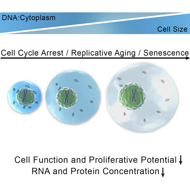
          Graphical Abstract

          Highlights

          • Deviation from normal cell size interferes with cell function and proliferation

          • DNA becomes limiting for cell function if cells grow too large

          • Uncoupling of protein synthesis and volume causes cytoplasm dilution in big cells

          • Excessive cell growth contributes to functional decline in senescence

          Abstract

          Optimal cell function requires maintenance of a narrow range of DNA:cytoplasm ratios and when cell size exceeds this ratio cytoplasmic dilution contributes to senescence

          Related collections

          Most cited references31

          • Record: found
          • Abstract: found
          • Article: not found

          Genomic expression programs in the response of yeast cells to environmental changes.

          We explored genomic expression patterns in the yeast Saccharomyces cerevisiae responding to diverse environmental transitions. DNA microarrays were used to measure changes in transcript levels over time for almost every yeast gene, as cells responded to temperature shocks, hydrogen peroxide, the superoxide-generating drug menadione, the sulfhydryl-oxidizing agent diamide, the disulfide-reducing agent dithiothreitol, hyper- and hypo-osmotic shock, amino acid starvation, nitrogen source depletion, and progression into stationary phase. A large set of genes (approximately 900) showed a similar drastic response to almost all of these environmental changes. Additional features of the genomic responses were specialized for specific conditions. Promoter analysis and subsequent characterization of the responses of mutant strains implicated the transcription factors Yap1p, as well as Msn2p and Msn4p, in mediating specific features of the transcriptional response, while the identification of novel sequence elements provided clues to novel regulators. Physiological themes in the genomic responses to specific environmental stresses provided insights into the effects of those stresses on the cell.
            Bookmark
            • Record: found
            • Abstract: found
            • Article: not found

            MultiNotch MS3 Enables Accurate, Sensitive, and Multiplexed Detection of Differential Expression across Cancer Cell Line Proteomes

            Multiplexed quantitation via isobaric chemical tags (e.g., tandem mass tags (TMT) and isobaric tags for relative and absolute quantitation (iTRAQ)) has the potential to revolutionize quantitative proteomics. However, until recently the utility of these tags was questionable due to reporter ion ratio distortion resulting from fragmentation of coisolated interfering species. These interfering signals can be negated through additional gas-phase manipulations (e.g., MS/MS/MS (MS3) and proton-transfer reactions (PTR)). These methods, however, have a significant sensitivity penalty. Using isolation waveforms with multiple frequency notches (i.e., synchronous precursor selection, SPS), we coisolated and cofragmented multiple MS2 fragment ions, thereby increasing the number of reporter ions in the MS3 spectrum 10-fold over the standard MS3 method (i.e., MultiNotch MS3). By increasing the reporter ion signals, this method improves the dynamic range of reporter ion quantitation, reduces reporter ion signal variance, and ultimately produces more high-quality quantitative measurements. To demonstrate utility, we analyzed biological triplicates of eight colon cancer cell lines using the MultiNotch MS3 method. Across all the replicates we quantified 8 378 proteins in union and 6 168 proteins in common. Taking into account that each of these quantified proteins contains eight distinct cell-line measurements, this data set encompasses 174 704 quantitative ratios each measured in triplicate across the biological replicates. Herein, we demonstrate that the MultiNotch MS3 method uniquely combines multiplexing capacity with quantitative sensitivity and accuracy, drastically increasing the informational value obtainable from proteomic experiments.
              Bookmark
              • Record: found
              • Abstract: found
              • Article: not found

              Increasing the multiplexing capacity of TMTs using reporter ion isotopologues with isobaric masses.

              Quantitative mass spectrometry methods offer near-comprehensive proteome coverage; however, these methods still suffer with regards to sample throughput. Multiplex quantitation via isobaric chemical tags (e.g., TMT and iTRAQ) provides an avenue for mass spectrometry-based proteome quantitation experiments to move away from simple binary comparisons and toward greater parallelization. Herein, we demonstrate a straightforward method for immediately expanding the throughput of the TMT isobaric reagents from 6-plex to 8-plex. This method is based upon our ability to resolve the isotopic shift that results from substituting a (15)N for a (13)C. In an accommodation to the preferred fragmentation pathways of ETD, the TMT-127 and -129 reagents were recently modified such that a (13)C was exchanged for a (15)N. As a result of this substitution, the new TMT reporter ions are 6.32 mDa lighter. Even though the mass difference between these reporter ion isotopologues is incredibly small, modern high-resolution and mass accuracy analyzers can resolve these ions. On the basis of our ability to resolve and accurately measure the relative intensity of these isobaric reporter ions, we demonstrate that we are able to quantify across eight samples simultaneously by combining the (13)C- and (15)N-containing reporter ions. Considering the structure of the TMT reporter ion, we believe this work serves as a blueprint for expanding the multiplexing capacity of the TMT reagents to at least 10-plex and possibly up to 18-plex.
                Bookmark

                Author and article information

                Contributors
                Journal
                Cell
                Cell
                Cell
                Cell Press
                0092-8674
                1097-4172
                21 February 2019
                21 February 2019
                : 176
                : 5
                : 1083-1097.e18
                Affiliations
                [1 ]David H. Koch Institute for Integrative Cancer Research, Howard Hughes Medical Institute, Massachusetts Institute of Technology, Cambridge, MA 02139, USA
                [2 ]Department of Biology, Massachusetts Institute of Technology, Cambridge, MA 02139, USA
                [3 ]Department of Systems Biology, Harvard Medical School, Boston, Massachusetts 02115, USA
                [4 ]Institute for Systems Genetics, New York University Langone Health, New York, NY 10016, USA
                [5 ]Cell Fate and Gene Regulation Laboratory, The Francis Crick Institute, 1 Midland Road, NW1 1AT London, UK
                [6 ]David H. Koch Institute for Integrative Cancer Research, Massachusetts Institute of Technology, Cambridge, MA 02139, USA
                [7 ]Department of Biological Engineering, Massachusetts Institute of Technology, Cambridge, MA 02139, USA
                [8 ]Department of Mechanical Engineering, Massachusetts Institute of Technology, Cambridge, MA 02139, USA
                [9 ]Department of Cell Biology, Harvard Medical School, Boston, MA 02115, USA
                [10 ]Department of Biological Chemistry and Molecular Pharmacology, Harvard Medical School, Boston, MA 02115, USA
                [11 ]MRC Laboratory for Molecular Cell Biology, University College London, Gower Street, London, WC1E 6BT, UK
                [12 ]Novartis Institute for Biomedical Research, Oncology Department, Cambridge, MA 02139
                Author notes
                []Corresponding author angelika@ 123456mit.edu
                [13]

                Lead Contact

                Article
                S0092-8674(19)30051-0
                10.1016/j.cell.2019.01.018
                6386581
                30739799
                7d4f8d5a-9ce2-4cdb-b44f-a7d83a1021c0
                © 2019 The Author(s)

                This is an open access article under the CC BY license (http://creativecommons.org/licenses/by/4.0/).

                History
                : 27 February 2018
                : 15 November 2018
                : 9 January 2019
                Categories
                Article

                Cell biology
                Cell biology

                Comments

                Comment on this article