22
views
0
recommends
+1 Recommend
0 collections
    0
    shares
      • Record: found
      • Abstract: found
      • Article: found
      Is Open Access

      Exosomes: Decreased Sensitivity of Lung Cancer A549 Cells to Cisplatin

      research-article

      Read this article at

      Bookmark
          There is no author summary for this article yet. Authors can add summaries to their articles on ScienceOpen to make them more accessible to a non-specialist audience.

          Abstract

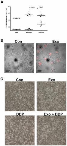
          Exosomes are small extracellular membrane vesicles of endocytic origin released by many cells that could be found in most body fluids. The main functions of exosomes are cellular communication and cellular waste clean-up. This study was conducted to determine the involvement of exosomes in the regulation of sensitivity of the lung cancer cell line A549 to cisplatin (DDP). When DDP was added to A549 cells, exosomes secretion was strengthened. Addition of the secreted exosomes to other A549 cells increased the resistance of these A549 cells to DDP. Upon exposure of A549 to DDP, the expression levels of several miRNAs and mRNAs reportedly associated with DDP sensitivity changed significantly in exosomes; these changes may mediate the resistance of A549 cells to DDP. Exosomes released by A549 cells during DDP exposure decreased the sensitivity of other A549 cells to DDP, which may be mediated by miRNAs and mRNAs exchange by exosomes via cell-to-cell communication. Although the detailed mechanism of resistance remains unclear, we believed that inhibition of exosomes formation and release might present a novel strategy for lung cancer treatment in the future.

          Related collections

          Most cited references18

          • Record: found
          • Abstract: found
          • Article: not found

          Exosome-mediated transfer of miR-133b from multipotent mesenchymal stromal cells to neural cells contributes to neurite outgrowth.

          Multipotent mesenchymal stromal cells (MSCs) have potential therapeutic benefit for the treatment of neurological diseases and injury. MSCs interact with and alter brain parenchymal cells by direct cell-cell communication and/or by indirect secretion of factors and thereby promote functional recovery. In this study, we found that MSC treatment of rats subjected to middle cerebral artery occlusion (MCAo) significantly increased microRNA 133b (miR-133b) level in the ipsilateral hemisphere. In vitro, miR-133b levels in MSCs and in their exosomes increased after MSCs were exposed to ipsilateral ischemic tissue extracts from rats subjected to MCAo. miR-133b levels were also increased in primary cultured neurons and astrocytes treated with the exosome-enriched fractions released from these MSCs. Knockdown of miR-133b in MSCs confirmed that the increased miR-133b level in astrocytes is attributed to their transfer from MSCs. Further verification of this exosome-mediated intercellular communication was performed using a cel-miR-67 luciferase reporter system and an MSC-astrocyte coculture model. Cel-miR-67 in MSCs was transferred to astrocytes via exosomes between 50 and 100 nm in diameter. Our data suggest that the cel-miR-67 released from MSCs was primarily contained in exosomes. A gap junction intercellular communication inhibitor arrested the exosomal microRNA communication by inhibiting exosome release. Cultured neurons treated with exosome-enriched fractions from MSCs exposed to 72 hours post-MCAo brain extracts significantly increased the neurite branch number and total neurite length. This study provides the first demonstration that MSCs communicate with brain parenchymal cells and may regulate neurite outgrowth by transfer of miR-133b to neural cells via exosomes. Copyright © 2012 AlphaMed Press.
            Bookmark
            • Record: found
            • Abstract: found
            • Article: not found

            Malignant effusions and immunogenic tumour-derived exosomes.

            Exosomes derived from tumours are small vesicles released in vitro by tumour cell lines in culture supernatants. To assess the role of these exosomes in vivo, we examined malignant effusions for their presence. We also investigated whether these exosomes could induce production of tumour-specific T cells when pulsed with dendritic cells. We isolated exosomes by ultracentrifugation on sucrose and D(2)O gradients of 11 malignant effusions. We characterised exosomes with Western blot analyses, immunoelectron microscopy, and in-vitro stimulations of autologous T lymphocytes. Malignant effusions accumulate high numbers of membrane vesicles that have a mean diameter of 80 nm (SD 30). These vesicles have antigen-presenting molecules (MHC class-I heat-shock proteins), tetraspanins (CD81), and tumour antigens (Her2/Neu, Mart1, TRP, gp100). These criteria, including their morphological characteristics, indicate the similarities between these vesicles and exosomes. Exosomes from patients with melanoma deliver Mart1 tumour antigens to dendritic cells derived from monocytes (MD-DCs) for cross presentation to clones of cytotoxic T lymphocytes specific to Mart1. In seven of nine patients with cancer, lymphocytes specific to the tumour could be efficiently expanded from peripheral blood cells by pulsing autologous MD-DCs with autologous ascitis exosomes. In one patient tested, we successfully expanded a restricted T-cell repertoire, which could not be recovered carcinomatosis nodules. Exosomes derived from tumours accumulate in ascites from patients with cancer. Ascitis exosomes are a natural and new source of tumour-rejection antigens, opening up new avenues for immunisation against cancers.
              Bookmark
              • Record: found
              • Abstract: found
              • Article: not found

              Abnormal lysosomal trafficking and enhanced exosomal export of cisplatin in drug-resistant human ovarian carcinoma cells.

              Previous work has shown that cisplatin (CDDP) becomes concentrated in lysosomes, and that acquired resistance to CDDP is associated with abnormalities of protein trafficking and secretion. The lysosomal compartment in CDDP-sensitive 2008 human ovarian carcinoma cells was compared with that in CDDP-resistant 2008/C13*5.25 subline using deconvoluting imaging and specific dyes and antibodies. The lysosomal compartment in CDDP-resistant cells was reduced to just 40% of that in the parental CDDP-sensitive cells (P<0.002). This was accompanied by a reduced expression of the lysosome-associated proteins 1 and 2 (LAMP1 and LAMP2) as determined by both microscopy and Western blot analysis. The CDDP-resistant cells released more protein as exosomes and Western blot analysis revealed that these exosomes contained substantially more LAMP1 than those released by the CDDP-sensitive cells. Following loading of the whole cell with CDDP, the exosomes released from 2008/C13*5.25 cells contained 2.6-fold more platinum than those released from sensitive cells. Enhanced exosomal export was accompanied by higher exosomal levels of the putative CDDP export transporters MRP2, ATP7A, and ATP7B. Expression profiling identified significant increases in the expression of several genes whose products function in membrane fusion and vesicle trafficking. This study shows that the lysosomal compartment of human ovarian carcinoma cells selected for stable resistance to CDDP is markedly reduced in size, and that these cells abnormally sort some lysosomal proteins and the putative CDDP transporters into an exosomal pathway that also exports CDDP.
                Bookmark

                Author and article information

                Contributors
                Role: Editor
                Journal
                PLoS One
                PLoS ONE
                plos
                plosone
                PLoS ONE
                Public Library of Science (San Francisco, USA )
                1932-6203
                2014
                21 February 2014
                : 9
                : 2
                : e89534
                Affiliations
                [1 ]Research Center for Clinical Oncology, Nanjing Medical University Affiliated Cancer Hospital, Jiangsu Cancer Hospital and Jiangsu Institute of Cancer Research, Nanjing, Jiangsu Province, China
                [2 ]Nanjing University of Technology, Nanjing, Jiangsu Province, China
                [3 ]Southeast University, Nanjing, Jiangsu Province, China
                Wayne State University School of Medicine, United States of America
                Author notes

                Competing Interests: The authors have declared that no competing interests exist.

                Conceived and designed the experiments: XX SY SL JW RW HC YZ JF. Performed the experiments: XX SY SL. Analyzed the data: XX SY JW RW HC JF. Contributed reagents/materials/analysis tools: XX SY YZ. Wrote the paper: XX SY SL JW JF.

                Article
                PONE-D-13-41080
                10.1371/journal.pone.0089534
                3931805
                24586853
                7d377ade-604d-428e-811e-dfbe4c440fef
                Copyright @ 2014

                This is an open-access article distributed under the terms of the Creative Commons Attribution License, which permits unrestricted use, distribution, and reproduction in any medium, provided the original author and source are credited.

                History
                : 8 October 2013
                : 22 January 2014
                Page count
                Pages: 6
                Funding
                This work was supported by the National Natural Science Foundation of China (No. 81372396) and the Six Talent Peak of Human Affairs Hall in Jiangsu Province (No. 40). The funders had no role in study design, data collection and analysis, decision to publish, or preparation of the manuscript.
                Categories
                Research Article
                Biology
                Molecular cell biology
                Cell death
                Cell growth
                Membranes and sorting
                Medicine
                Drugs and devices
                Oncology
                Cancer treatment
                Chemotherapy and drug treatment
                Cancers and neoplasms
                Lung and intrathoracic tumors
                Adenocarcinoma of the lung
                Non-small cell lung cancer

                Uncategorized
                Uncategorized

                Comments

                Comment on this article