2
views
0
recommends
+1 Recommend
0 collections
    0
    shares
      • Record: found
      • Abstract: found
      • Article: found
      Is Open Access

      RiFNet: Automated rib fracture detection in postmortem computed tomography

      research-article

      Read this article at

      Bookmark
          There is no author summary for this article yet. Authors can add summaries to their articles on ScienceOpen to make them more accessible to a non-specialist audience.

          Abstract

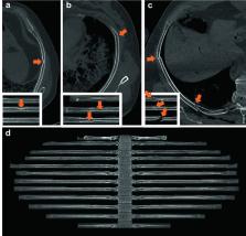
          Imaging techniques are widely used for medical diagnostics. In some cases, a lack of medical practitioners who can manually analyze the images can lead to a bottleneck. Consequently, we developed a custom-made convolutional neural network (RiFNet =  Rib Fracture Network) that can detect rib fractures in postmortem computed tomography. In a retrospective cohort study, we retrieved PMCT data from 195 postmortem cases with rib fractures from July 2017 to April 2018 from our database. The computed tomography data were prepared using a plugin in the commercial imaging software Syngo.via whereby the rib cage was unfolded on a single-in-plane image reformation. Out of the 195 cases, a total of 585 images were extracted and divided into two groups labeled “with” and “without” fractures. These two groups were subsequently divided into training, validation, and test datasets to assess the performance of RiFNet. In addition, we explored the possibility of applying transfer learning techniques on our dataset by choosing two independent noncommercial off-the-shelf convolutional neural network architectures (ResNet50 V2 and Inception V3) and compared the performances of those two with RiFNet. When using pre-trained convolutional neural networks, we achieved an F 1 score of 0.64 with Inception V3 and an F 1 score of 0.61 with ResNet50 V2. We obtained an average F 1 score of 0.91 ± 0.04 with RiFNet. RiFNet is efficient in detecting rib fractures on postmortem computed tomography. Transfer learning techniques are not necessarily well adapted to make classifications in postmortem computed tomography.

          Supplementary Information

          The online version contains supplementary material available at 10.1007/s12024-021-00431-8.

          Related collections

          Most cited references24

          • Record: found
          • Abstract: found
          • Article: not found

          Deep learning.

          Deep learning allows computational models that are composed of multiple processing layers to learn representations of data with multiple levels of abstraction. These methods have dramatically improved the state-of-the-art in speech recognition, visual object recognition, object detection and many other domains such as drug discovery and genomics. Deep learning discovers intricate structure in large data sets by using the backpropagation algorithm to indicate how a machine should change its internal parameters that are used to compute the representation in each layer from the representation in the previous layer. Deep convolutional nets have brought about breakthroughs in processing images, video, speech and audio, whereas recurrent nets have shone light on sequential data such as text and speech.
            Bookmark
            • Record: found
            • Abstract: not found
            • Article: not found

            Backpropagation Applied to Handwritten Zip Code Recognition

              Bookmark
              • Record: found
              • Abstract: found
              • Article: not found

              Efficient multi-scale 3D CNN with fully connected CRF for accurate brain lesion segmentation

              We propose a dual pathway, 11-layers deep, three-dimensional Convolutional Neural Network for the challenging task of brain lesion segmentation. The devised architecture is the result of an in-depth analysis of the limitations of current networks proposed for similar applications. To overcome the computational burden of processing 3D medical scans, we have devised an efficient and effective dense training scheme which joins the processing of adjacent image patches into one pass through the network while automatically adapting to the inherent class imbalance present in the data. Further, we analyze the development of deeper, thus more discriminative 3D CNNs. In order to incorporate both local and larger contextual information, we employ a dual pathway architecture that processes the input images at multiple scales simultaneously. For post-processing of the network's soft segmentation, we use a 3D fully connected Conditional Random Field which effectively removes false positives. Our pipeline is extensively evaluated on three challenging tasks of lesion segmentation in multi-channel MRI patient data with traumatic brain injuries, brain tumours, and ischemic stroke. We improve on the state-of-the-art for all three applications, with top ranking performance on the public benchmarks BRATS 2015 and ISLES 2015. Our method is computationally efficient, which allows its adoption in a variety of research and clinical settings. The source code of our implementation is made publicly available.
                Bookmark

                Author and article information

                Contributors
                Akos.Dobay@uzh.ch
                Journal
                Forensic Sci Med Pathol
                Forensic Sci Med Pathol
                Forensic Science, Medicine, and Pathology
                Springer US (New York )
                1547-769X
                1556-2891
                28 October 2021
                28 October 2021
                2022
                : 18
                : 1
                : 20-29
                Affiliations
                [1 ]GRID grid.7400.3, ISNI 0000 0004 1937 0650, Zurich Institute of Forensic Medicine, , University of Zurich, ; Winterthurerstrasse 190/52, CH-8057 Zurich, Switzerland
                [2 ]GRID grid.267207.6, ISNI 0000 0001 2218 5518, Department of Mathematics, , University of St. Thomas, ; St. Paul, Minnesota, 55105-1079 USA
                [3 ]GRID grid.412004.3, ISNI 0000 0004 0478 9977, Institute of Diagnostic and Interventional Radiology, , University Hospital Zurich, ; Rämistrasse 100, 8091 Zurich, Switzerland
                Author information
                http://orcid.org/0000-0001-6492-9298
                Article
                431
                10.1007/s12024-021-00431-8
                8921053
                34709561
                7d02a11a-3833-402c-8b82-652c83acf335
                © The Author(s) 2021

                Open AccessThis article is licensed under a Creative Commons Attribution 4.0 International License, which permits use, sharing, adaptation, distribution and reproduction in any medium or format, as long as you give appropriate credit to the original author(s) and the source, provide a link to the Creative Commons licence, and indicate if changes were made. The images or other third party material in this article are included in the article's Creative Commons licence, unless indicated otherwise in a credit line to the material. If material is not included in the article's Creative Commons licence and your intended use is not permitted by statutory regulation or exceeds the permitted use, you will need to obtain permission directly from the copyright holder. To view a copy of this licence, visit http://creativecommons.org/licenses/by/4.0/.

                History
                : 20 September 2021
                Funding
                Funded by: University of Zurich
                Categories
                Original Article
                Custom metadata
                © Springer Science+Business Media, LLC, part of Springer Nature 2022

                Forensic science
                deep learning,convolutional neural networks,computed tomography,forensic sciences,pmct

                Comments

                Comment on this article