0
views
0
recommends
+1 Recommend
0 collections
    0
    shares
      • Record: found
      • Abstract: found
      • Article: found
      Is Open Access

      Automated evaluation of retinal hyperreflective foci changes in diabetic macular edema patients before and after intravitreal injection

      research-article

      Read this article at

      Bookmark
          There is no author summary for this article yet. Authors can add summaries to their articles on ScienceOpen to make them more accessible to a non-specialist audience.

          Abstract

          Purpose

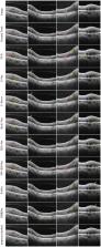
          Fast and automated reconstruction of retinal hyperreflective foci (HRF) is of great importance for many eye-related disease understanding. In this paper, we introduced a new automated framework, driven by recent advances in deep learning to automatically extract 12 three-dimensional parameters from the segmented hyperreflective foci in optical coherence tomography (OCT).

          Methods

          Unlike traditional convolutional neural networks, which struggle with long-range feature correlations, we introduce a spatial and channel attention module within the bottleneck layer, integrated into the nnU-Net architecture. Spatial Attention Block aggregates features across spatial locations to capture related features, while Channel Attention Block heightens channel feature contrasts. The proposed model was trained and tested on 162 retinal OCT volumes of patients with diabetic macular edema (DME), yielding robust segmentation outcomes. We further investigate HRF’s potential as a biomarker of DME.

          Results

          Results unveil notable discrepancies in the amount and volume of HRF subtypes. In the whole retinal layer (WR), the mean distance from HRF to the retinal pigmented epithelium was significantly reduced after treatment. In WR, the improvement in central macular thickness resulting from intravitreal injection treatment was positively correlated with the mean distance from HRF subtypes to the fovea.

          Conclusion

          Our study demonstrates the applicability of OCT for automated quantification of retinal HRF in DME patients, offering an objective, quantitative approach for clinical and research applications.

          Related collections

          Most cited references44

          • Record: found
          • Abstract: not found
          • Book Chapter: not found

          U-Net: Convolutional Networks for Biomedical Image Segmentation

            Bookmark
            • Record: found
            • Abstract: found
            • Article: found
            Is Open Access

            Global Prevalence and Major Risk Factors of Diabetic Retinopathy

            OBJECTIVE To examine the global prevalence and major risk factors for diabetic retinopathy (DR) and vision-threatening diabetic retinopathy (VTDR) among people with diabetes. RESEARCH DESIGN AND METHODS A pooled analysis using individual participant data from population-based studies around the world was performed. A systematic literature review was conducted to identify all population-based studies in general populations or individuals with diabetes who had ascertained DR from retinal photographs. Studies provided data for DR end points, including any DR, proliferative DR, diabetic macular edema, and VTDR, and also major systemic risk factors. Pooled prevalence estimates were directly age-standardized to the 2010 World Diabetes Population aged 20–79 years. RESULTS A total of 35 studies (1980–2008) provided data from 22,896 individuals with diabetes. The overall prevalence was 34.6% (95% CI 34.5–34.8) for any DR, 6.96% (6.87–7.04) for proliferative DR, 6.81% (6.74–6.89) for diabetic macular edema, and 10.2% (10.1–10.3) for VTDR. All DR prevalence end points increased with diabetes duration, hemoglobin A1c, and blood pressure levels and were higher in people with type 1 compared with type 2 diabetes. CONCLUSIONS There are approximately 93 million people with DR, 17 million with proliferative DR, 21 million with diabetic macular edema, and 28 million with VTDR worldwide. Longer diabetes duration and poorer glycemic and blood pressure control are strongly associated with DR. These data highlight the substantial worldwide public health burden of DR and the importance of modifiable risk factors in its occurrence. This study is limited by data pooled from studies at different time points, with different methodologies and population characteristics.
              Bookmark
              • Record: found
              • Abstract: found
              • Article: not found

              nnU-Net: a self-configuring method for deep learning-based biomedical image segmentation

              Biomedical imaging is a driver of scientific discovery and a core component of medical care and is being stimulated by the field of deep learning. While semantic segmentation algorithms enable image analysis and quantification in many applications, the design of respective specialized solutions is non-trivial and highly dependent on dataset properties and hardware conditions. We developed nnU-Net, a deep learning-based segmentation method that automatically configures itself, including preprocessing, network architecture, training and post-processing for any new task. The key design choices in this process are modeled as a set of fixed parameters, interdependent rules and empirical decisions. Without manual intervention, nnU-Net surpasses most existing approaches, including highly specialized solutions on 23 public datasets used in international biomedical segmentation competitions. We make nnU-Net publicly available as an out-of-the-box tool, rendering state-of-the-art segmentation accessible to a broad audience by requiring neither expert knowledge nor computing resources beyond standard network training.
                Bookmark

                Author and article information

                Contributors
                Role: Role: Role: Role: Role: Role:
                URI : https://loop.frontiersin.org/people/2188854/overviewRole: Role: Role: Role:
                URI : https://loop.frontiersin.org/people/1409473/overviewRole:
                URI : https://loop.frontiersin.org/people/1074083/overviewRole:
                Role: Role:
                URI : https://loop.frontiersin.org/people/2423307/overviewRole: Role:
                Role: Role:
                Role: Role: Role:
                URI : https://loop.frontiersin.org/people/2152900/overviewRole: Role: Role:
                URI : https://loop.frontiersin.org/people/1362172/overviewRole: Role: Role: Role: Role: Role: Role: Role: Role: Role: Role:
                Journal
                Front Med (Lausanne)
                Front Med (Lausanne)
                Front. Med.
                Frontiers in Medicine
                Frontiers Media S.A.
                2296-858X
                06 October 2023
                2023
                : 10
                : 1280714
                Affiliations
                [1] 1Cixi Biomedical Research Institute, Wenzhou Medical University , Ningbo, China
                [2] 2Institute of Biomedical Engineering, Ningbo Institute of Materials Technology and Engineering, Chinese Academy of Sciences , Ningbo, China
                [3] 3The Affiliated Ningbo Eye Hospital of Wenzhou Medical University , Ningbo, China
                [4] 4Department of Neurology, West China Hospital, Sichuan University , Chengdu, China
                [5] 5Health Science Center, Ningbo University , Ningbo, China
                Author notes

                Edited by: Meng Wang, Agency for Science, Technology and Research (A*STAR), Singapore

                Reviewed by: Yi Zhou, Soochow University, China; Ao Cheng, Soochow University, China

                *Correspondence: Yitian Zhao, yitian.zhao@ 123456nimte.ac.cn

                These authors have contributed equally to this work

                Article
                10.3389/fmed.2023.1280714
                10587607
                37869163
                7bf8ff27-a5d2-44dd-9341-009c45f4bf21
                Copyright © 2023 Wang, Zhang, Ma, Kwapong, Ying, Lu, Ma, Yan, Yi and Zhao.

                This is an open-access article distributed under the terms of the Creative Commons Attribution License (CC BY). The use, distribution or reproduction in other forums is permitted, provided the original author(s) and the copyright owner(s) are credited and that the original publication in this journal is cited, in accordance with accepted academic practice. No use, distribution or reproduction is permitted which does not comply with these terms.

                History
                : 21 August 2023
                : 21 September 2023
                Page count
                Figures: 4, Tables: 6, Equations: 9, References: 46, Pages: 13, Words: 9334
                Funding
                The author(s) declare financial support was received for the research, authorship, and/or publication of this article. This work was supported in part by the National Natural Science Foundation of China (62272444); Zhejiang Provincial Natural Science Foundation (LR22F020008); Youth Innovation Promotion Association CAS (2021298). National Science Foundation Program of China (62302488); Zhejiang Provincial Natural Science Foundation of China (LQ23F010007).
                Categories
                Medicine
                Original Research
                Custom metadata
                Ophthalmology

                diabetic macular edema,hyperreflective foci,optical coherence tomography,artificial intelligence,deep learning

                Comments

                Comment on this article