7
views
0
recommends
+1 Recommend
0 collections
    0
    shares
      • Record: found
      • Abstract: found
      • Article: found
      Is Open Access

      Hyper-Reflecting Foci in Multiple Sclerosis Retina Associate With Macrophage/Microglia-Derived Cytokines in Cerebrospinal Fluid

      research-article

      Read this article at

      Bookmark
          There is no author summary for this article yet. Authors can add summaries to their articles on ScienceOpen to make them more accessible to a non-specialist audience.

          Abstract

          Background

          Increasing evidence suggests that retinal hyper-reflecting foci (HRF) might be clusters of activated and proliferating microglia. Since microglia are widespread activated in multiple sclerosis (MS) brain, its evaluation in retina may help to understand and monitor MS-related pathology.

          Aim

          This study aims at investigating the association of HRF with cerebrospinal fluid (CSF) cytokines and MRI parameters in relapsing–remitting MS (RRMS).

          Methods

          Nineteen RRMS at clinical onset and 15 non-inflammatory neurological disorders (NIND) underwent brain 3T MRI and CSF examination. Optical coherence tomography (OCT) analysis, including HRF count, was performed on RRMS patients. Sixty-nine cytokines/chemokines were analyzed in the CSF by multiplex technology.

          Results

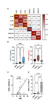
          In RRMS, HRF count in the ganglion cell layer (GCL) was associated with IL-1Ra, IL-9, IL-15, IFN-γ, and G-CSF. Moreover, in RRMS patients CSF concentrations of IL-1Ra and G-CSF associated with global cortical thickness. The HRF count in the inner nuclear layer (INL) correlated with IL-22, IL-34, IL-35, CXCL-2, CXCL-10, and CXCL-13, and multivariate analysis confirmed a strong association (r 2: 0.47) with both CXCL-2 (β: -0.965, p = 0.0052) and CXCL-13 (β: 0.241, p = 0.018). This latter cytokine increased in RRMS with high HRF count compared with NIND and RRMS with low HRF count. Finally, the CXCL-13/CXCL-2 ratio strongly associated with HRF count (r: 0.8, p < 0.005) and cortical lesion volume (r: 0.5, p < 0.05).

          Conclusions

          The association of HRF with intrathecally produced monocyte/microglia-derived cytokines confirms their microglial origin and indicates they are worth further evaluating as markers of activated microglia.

          Related collections

          Most cited references36

          • Record: found
          • Abstract: found
          • Article: found

          Diagnosis of multiple sclerosis: 2017 revisions of the McDonald criteria

          The 2010 McDonald criteria for the diagnosis of multiple sclerosis are widely used in research and clinical practice. Scientific advances in the past 7 years suggest that they might no longer provide the most up-to-date guidance for clinicians and researchers. The International Panel on Diagnosis of Multiple Sclerosis reviewed the 2010 McDonald criteria and recommended revisions. The 2017 McDonald criteria continue to apply primarily to patients experiencing a typical clinically isolated syndrome, define what is needed to fulfil dissemination in time and space of lesions in the CNS, and stress the need for no better explanation for the presentation. The following changes were made: in patients with a typical clinically isolated syndrome and clinical or MRI demonstration of dissemination in space, the presence of CSF-specific oligoclonal bands allows a diagnosis of multiple sclerosis; symptomatic lesions can be used to demonstrate dissemination in space or time in patients with supratentorial, infratentorial, or spinal cord syndrome; and cortical lesions can be used to demonstrate dissemination in space. Research to further refine the criteria should focus on optic nerve involvement, validation in diverse populations, and incorporation of advanced imaging, neurophysiological, and body fluid markers.
            Bookmark
            • Record: found
            • Abstract: found
            • Article: not found

            N4ITK: improved N3 bias correction.

            A variant of the popular nonparametric nonuniform intensity normalization (N3) algorithm is proposed for bias field correction. Given the superb performance of N3 and its public availability, it has been the subject of several evaluation studies. These studies have demonstrated the importance of certain parameters associated with the B-spline least-squares fitting. We propose the substitution of a recently developed fast and robust B-spline approximation routine and a modified hierarchical optimization scheme for improved bias field correction over the original N3 algorithm. Similar to the N3 algorithm, we also make the source code, testing, and technical documentation of our contribution, which we denote as "N4ITK," available to the public through the Insight Toolkit of the National Institutes of Health. Performance assessment is demonstrated using simulated data from the publicly available Brainweb database, hyperpolarized (3)He lung image data, and 9.4T postmortem hippocampus data.
              Bookmark
              • Record: found
              • Abstract: found
              • Article: not found

              Fast robust automated brain extraction.

              An automated method for segmenting magnetic resonance head images into brain and non-brain has been developed. It is very robust and accurate and has been tested on thousands of data sets from a wide variety of scanners and taken with a wide variety of MR sequences. The method, Brain Extraction Tool (BET), uses a deformable model that evolves to fit the brain's surface by the application of a set of locally adaptive model forces. The method is very fast and requires no preregistration or other pre-processing before being applied. We describe the new method and give examples of results and the results of extensive quantitative testing against "gold-standard" hand segmentations, and two other popular automated methods. Copyright 2002 Wiley-Liss, Inc.
                Bookmark

                Author and article information

                Contributors
                Journal
                Front Immunol
                Front Immunol
                Front. Immunol.
                Frontiers in Immunology
                Frontiers Media S.A.
                1664-3224
                19 May 2022
                2022
                : 13
                : 852183
                Affiliations
                [1] 1 Multiple Sclerosis Centre, Neurology Clinic, Department of Neuroscience, Università degli Studi di Padova , Padova, Italy
                [2] 2 Ophthalmology Clinic, Department of Neuroscience, Università degli Studi di Padova , Padova, Italy
                Author notes

                Edited by: Kin-Sang Cho, Schepens Eye Research Institute, Harvard Medical School, United States

                Reviewed by: Frederike Cosima Oertel, University of California, San Francisco, United States; Shiv Saidha, Johns Hopkins Medicine, United States

                *Correspondence: Marco Puthenparampil, marco.puthenparampil@ 123456unipd.it

                This article was submitted to Multiple Sclerosis and Neuroimmunology, a section of the journal Frontiers in Immunology

                Article
                10.3389/fimmu.2022.852183
                9160385
                35664007
                14d1eb6d-bdea-412d-97c9-aee03159dae1
                Copyright © 2022 Puthenparampil, Torresin, Franciotta, Marin, De Napoli, Mauceri, Miante, Pilotto, Midena and Gallo

                This is an open-access article distributed under the terms of the Creative Commons Attribution License (CC BY). The use, distribution or reproduction in other forums is permitted, provided the original author(s) and the copyright owner(s) are credited and that the original publication in this journal is cited, in accordance with accepted academic practice. No use, distribution or reproduction is permitted which does not comply with these terms.

                History
                : 10 January 2022
                : 22 March 2022
                Page count
                Figures: 2, Tables: 3, Equations: 0, References: 37, Pages: 8, Words: 4199
                Categories
                Immunology
                Original Research

                Immunology
                multiple sclerosis,oct,microglia,retina,cerebrospinal fluid,hyper-reflecting foci
                Immunology
                multiple sclerosis, oct, microglia, retina, cerebrospinal fluid, hyper-reflecting foci

                Comments

                Comment on this article