8
views
0
recommends
+1 Recommend
0 collections
    0
    shares
      • Record: found
      • Abstract: found
      • Article: found
      Is Open Access

      Imaging Hyperreflective Foci as an Inflammatory Biomarker after Anti-VEGF Treatment in Neovascular Age-Related Macular Degeneration Patients with Optical Coherence Tomography Angiography

      research-article

      Read this article at

      Bookmark
          There is no author summary for this article yet. Authors can add summaries to their articles on ScienceOpen to make them more accessible to a non-specialist audience.

          Abstract

          Purpose

          To investigate the hyperreflective foci (HRF) as an inflammatory biomarker using optical coherence tomography angiography (OCTA) in neovascular age-related macular degeneration (AMD) patients after antivascular endothelial growth factor (anti-VEGF) treatment and its association with the retinal microcapillary density.

          Methods

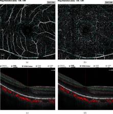
          Twenty-five eyes from 25 patients with neovascular AMD were included in the study. All eyes were imaged with OCTA at baseline (M0) and after 3 consecutive injections (M3; injection performed each month) of anti-VEGF. The number of HRF in the superficial capillary plexus (SCP), deep capillary plexus (DCP), and outer retina was counted. The vascular density of the fovea, parafovea, and the whole macula, as well as the area of the foveal avascular zone (FAZ), was measured.

          Results

          The mean interval between baseline and follow-up with OCTA was 93.08 ± 5.00 (range, 85-101) days. Compared with the baseline, the number of HRF significantly decreased in DCP (7.52 ± 3.06 vs. 3.76 ± 1.48, P < 0.01) and outer retina (12.04 ± 4.91 vs. 5.88 ± 3.32, P < 0.01) after treatment. There was no significant difference for HRF number in the SCP, the vascular density (containing foveal, parafoveal, and whole macular), and FAZ area before and after treatments.

          Conclusion

          The number of HRF in DCP and outer retina might serve as an inflammatory biomarker in patients with neovascular AMD. The reduced HRF possibly represents the alleviation of inflammation after anti-VEGF treatment in patients with AMD.

          Related collections

          Most cited references31

          • Record: found
          • Abstract: found
          • Article: not found
          Is Open Access

          Global prevalence of age-related macular degeneration and disease burden projection for 2020 and 2040: a systematic review and meta-analysis.

          Numerous population-based studies of age-related macular degeneration have been reported around the world, with the results of some studies suggesting racial or ethnic differences in disease prevalence. Integrating these resources to provide summarised data to establish worldwide prevalence and to project the number of people with age-related macular degeneration from 2020 to 2040 would be a useful guide for global strategies. We did a systematic literature review to identify all population-based studies of age-related macular degeneration published before May, 2013. Only studies using retinal photographs and standardised grading classifications (the Wisconsin age-related maculopathy grading system, the international classification for age-related macular degeneration, or the Rotterdam staging system) were included. Hierarchical Bayesian approaches were used to estimate the pooled prevalence, the 95% credible intervals (CrI), and to examine the difference in prevalence by ethnicity (European, African, Hispanic, Asian) and region (Africa, Asia, Europe, Latin America and the Caribbean, North America, and Oceania). UN World Population Prospects were used to project the number of people affected in 2014 and 2040. Bayes factor was calculated as a measure of statistical evidence, with a score above three indicating substantial evidence. Analysis of 129,664 individuals (aged 30-97 years), with 12,727 cases from 39 studies, showed the pooled prevalence (mapped to an age range of 45-85 years) of early, late, and any age-related macular degeneration to be 8.01% (95% CrI 3.98-15.49), 0.37% (0.18-0.77), and 8.69% (4.26-17.40), respectively. We found a higher prevalence of early and any age-related macular degeneration in Europeans than in Asians (early: 11.2% vs 6.8%, Bayes factor 3.9; any: 12.3% vs 7.4%, Bayes factor 4.3), and early, late, and any age-related macular degeneration to be more prevalent in Europeans than in Africans (early: 11.2% vs 7.1%, Bayes factor 12.2; late: 0.5% vs 0.3%, 3.7; any: 12.3% vs 7.5%, 31.3). There was no difference in prevalence between Asians and Africans (all Bayes factors <1). Europeans had a higher prevalence of geographic atrophy subtype (1.11%, 95% CrI 0.53-2.08) than Africans (0.14%, 0.04-0.45), Asians (0.21%, 0.04-0.87), and Hispanics (0.16%, 0.05-0.46). Between geographical regions, cases of early and any age-related macular degeneration were less prevalent in Asia than in Europe and North America (early: 6.3% vs 14.3% and 12.8% [Bayes factor 2.3 and 7.6]; any: 6.9% vs 18.3% and 14.3% [3.0 and 3.8]). No significant gender effect was noted in prevalence (Bayes factor <1.0). The projected number of people with age-related macular degeneration in 2020 is 196 million (95% CrI 140-261), increasing to 288 million in 2040 (205-399). These estimates indicate the substantial global burden of age-related macular degeneration. Summarised data provide information for understanding the effect of the condition and provide data towards designing eye-care strategies and health services around the world. National Medical Research Council, Singapore. Copyright © 2014 Wong et al. Open Access article distributed under the terms of CC BY-NC-ND. Published by .. All rights reserved.
            Bookmark
            • Record: found
            • Abstract: found
            • Article: not found

            Microglia in neurodegenerative disease.

            Microglia, the resident macrophages of the CNS, are exquisitely sensitive to brain injury and disease, altering their morphology and phenotype to adopt a so-called activated state in response to pathophysiological brain insults. Morphologically activated microglia, like other tissue macrophages, exist as many different phenotypes, depending on the nature of the tissue injury. Microglial responsiveness to injury suggests that these cells have the potential to act as diagnostic markers of disease onset or progression, and could contribute to the outcome of neurodegenerative diseases. The persistence of activated microglia long after acute injury and in chronic disease suggests that these cells have an innate immune memory of tissue injury and degeneration. Microglial phenotype is also modified by systemic infection or inflammation. Evidence from some preclinical models shows that systemic manipulations can ameliorate disease progression, although data from other models indicates that systemic inflammation exacerbates disease progression. Systemic inflammation is associated with a decline in function in patients with chronic neurodegenerative disease, both acutely and in the long term. The fact that diseases with a chronic systemic inflammatory component are risk factors for Alzheimer disease implies that crosstalk occurs between systemic inflammation and microglia in the CNS.
              Bookmark
              • Record: found
              • Abstract: found
              • Article: not found

              Retinal microglia: just bystander or target for therapy?

              Resident microglial cells can be regarded as the immunological watchdogs of the brain and the retina. They are active sensors of their neuronal microenvironment and rapidly respond to various insults with a morphological and functional transformation into reactive phagocytes. There is strong evidence from animal models and in situ analyses of human tissue that microglial reactivity is a common hallmark of various retinal degenerative and inflammatory diseases. These include rare hereditary retinopathies such as retinitis pigmentosa and X-linked juvenile retinoschisis but also comprise more common multifactorial retinal diseases such as age-related macular degeneration, diabetic retinopathy, glaucoma, and uveitis as well as neurological disorders with ocular manifestation. In this review, we describe how microglial function is kept in balance under normal conditions by cross-talk with other retinal cells and summarize how microglia respond to different forms of retinal injury. In addition, we present the concept that microglia play a key role in local regulation of complement in the retina and specify aspects of microglial aging relevant for chronic inflammatory processes in the retina. We conclude that this resident immune cell of the retina cannot be simply regarded as bystander of disease but may instead be a potential therapeutic target to be modulated in the treatment of degenerative and inflammatory diseases of the retina.
                Bookmark

                Author and article information

                Contributors
                Journal
                Biomed Res Int
                Biomed Res Int
                BMRI
                BioMed Research International
                Hindawi
                2314-6133
                2314-6141
                2021
                3 February 2021
                : 2021
                : 6648191
                Affiliations
                1Department of Ophthalmology, Shanghai Tenth People's Hospital, Tongji University School of Medicine, Shanghai, China
                2Department of Ophthalmology, Shanghai General Hospital (Shanghai First People's Hospital), Shanghai Jiao Tong University, Shanghai, China
                3National Clinical Research Center for Eye Diseases, Shanghai Key Laboratory of Ocular Fundus Diseases, Shanghai Engineering Center for Visual Science and Photomedicine, Shanghai Engineering Center for Precise Diagnosis and Treatment of Eye Diseases, Shanghai, China
                4Tongji Eye Institute, Tongji University School of Medicine, Shanghai, China
                Author notes

                Academic Editor: Qian Wang

                Author information
                https://orcid.org/0000-0002-7829-944X
                https://orcid.org/0000-0002-9450-1812
                https://orcid.org/0000-0002-5671-8412
                https://orcid.org/0000-0001-5950-6573
                https://orcid.org/0000-0003-3378-2363
                https://orcid.org/0000-0002-8383-892X
                https://orcid.org/0000-0002-0115-7160
                https://orcid.org/0000-0003-2844-2225
                https://orcid.org/0000-0003-0601-4342
                Article
                10.1155/2021/6648191
                7878069
                33614783
                1ac0f518-ab3e-42dc-b9ea-6012f31cff49
                Copyright © 2021 Jing Wu et al.

                This is an open access article distributed under the Creative Commons Attribution License, which permits unrestricted use, distribution, and reproduction in any medium, provided the original work is properly cited.

                History
                : 30 December 2020
                : 20 January 2021
                : 27 January 2021
                Funding
                Funded by: Clinical Research and Cultivation Project of Shanghai Municipal Hospital, China
                Award ID: SHDC12019X30
                Funded by: Science and Technology Commission of Shanghai Municipality
                Award ID: 20142203200
                Award ID: 19495800700
                Funded by: National Natural Science Foundation of China
                Award ID: 81970811
                Award ID: 81970810
                Award ID: 81870667
                Award ID: 81570852
                Categories
                Research Article

                Comments

                Comment on this article