11
views
0
recommends
+1 Recommend
0 collections
    0
    shares
      • Record: found
      • Abstract: found
      • Article: found
      Is Open Access

      Indolent Infection After Lumbar Interbody Fusion: An Under-recognized Cause of Pseudarthrosis, Which Can Be Successfully Treated With Anterior Revision Fusion

      research-article
      , MD, , ScB, , ScB, , MD, , MD
      JAAOS Global Research & Reviews
      Wolters Kluwer

      Read this article at

      Bookmark
          There is no author summary for this article yet. Authors can add summaries to their articles on ScienceOpen to make them more accessible to a non-specialist audience.

          Introduction:

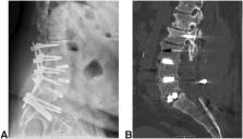
          Bacterial infection is a common etiology for pseudarthrosis after transforaminal lumbar interbody fusion, although it is often difficult to identify because of a delayed presentation and normal laboratory values. The primary goal of this study was to present a series of cases demonstrating patients with infection-related pseudarthrosis successfully managed with anterior revision.

          Methods:

          We retrospectively reviewed patients presenting to a single academic spine center who were found to have evidence of Cutibacterium acnes or coagulase-negative Staphylococcus infection on routine culturing of lumbar interbody fusion revisions from July 2019 to January 2021. All patients underwent salvage of a transforaminal lumbar interbody fusion pseudarthrosis through an anterior lumbar approach.

          Results:

          A total of six patients managed for pseudarthrosis secondary to suspected infection were eligible for this study (mean age 64.8 years, range 54-70 years; mean body mass index, range 24.5-39.1). Persistent radiculopathy was the primary presenting symptom in all patients with a mean time to revision of 17 months. Coagulase-negative Staphylococcus was the primary pathogen, identified from intraoperative samples in 50% of the cases. All patients demonstrated a resolution of symptoms after placement of an anterior lumbar interbody cage, without intraoperative complications, and a subsequent antibiotic regimen.

          Discussion:

          Indolent infection is an under-recognized cause of pseudarthrosis of the lumbar spine. Revision surgery through an anterior lumbar approach, which promotes ease of cage removal and optimized alignment and surface area available for revision fusion, is sufficient to manage pseudarthrosis due to infection.

          Related collections

          Most cited references32

          • Record: found
          • Abstract: not found
          • Article: not found

          Transforaminal lumbar interbody fusion (TLIF) versus posterior lumbar interbody fusion (PLIF) in lumbar spondylolisthesis: a systematic review and meta-analysis

            Bookmark
            • Record: found
            • Abstract: found
            • Article: not found

            Propionibacterium acnes infection after shoulder arthroplasty: a diagnostic challenge.

            This study reviewed a series of patients diagnosed with Propionibacterium acnes infection after shoulder arthroplasty in order to describe its clinical presentation, the means of diagnosis, and provide options for treatment. From 2002 to 2006, 11 patients diagnosed with P acnes infection after shoulder arthroplasty were retrospectively reviewed and analyzed for (1) clinical diagnosis; (2) laboratory data, including white blood cell count, erythrocyte sedimentation rate (ESR), and C-reactive protein (CRP); (3) fever; (4) number of days for laboratory growth of P acnes; (5) organism sensitivities; (6) antibiotic regimen and length of treatment; and (7) surgical management. Infection was diagnosed by 2 positive cultures. Five patients had an initial diagnosis of infection and underwent implant removal, placement of an antibiotic spacer, and staged reimplantation after a course of intravenous antibiotics. In the remaining 6 patients, surgical treatment varied according to the clinical diagnosis. When infection was recognized by intraoperative cultures, antibiotics were initiated. The average initial ESR and CRP values were 33 mm/h and 2 mg/dL, respectively. The average number of days from collection to a positive culture was 9. All cultures were sensitive to penicillin and clindamycin and universally resistant to metronidazole. Prosthetic joint infection secondary to P acnes is relatively rare; yet, when present, is an important cause of clinical implant failure. Successful treatment is hampered because clinical findings may be subtle, many of the traditional signs of infection are not present, and cultures may not be positive for as long as 2 weeks. Copyright 2010 Journal of Shoulder and Elbow Surgery Board of Trustees. Published by Mosby, Inc. All rights reserved.
              Bookmark
              • Record: found
              • Abstract: found
              • Article: not found

              Lumbar pseudarthrosis: a review of current diagnosis and treatment.

              OBJECT Failed solid bony fusion, or pseudarthrosis, is a well-known complication of lumbar arthrodesis. Recent advances in radiographic technology, biologics, instrumentation, surgical technique, and understanding of the local biology have all aided in the prevention and treatment of pseudarthrosis. Here, the current literature on the diagnosis and management of lumbar pseudarthroses is reviewed. METHODS A systematic literature review was conducted using the MEDLINE and Embase databases in order to search for the current radiographie diagnosis and surgical treatment methods published in the literature (1985 to present). Inclusion criteria included: 1) published in English; 2) level of evidence I-III; 3) diagnosis of degenerative lumbar spine conditions and/or history of lumbar spine fusion surgery; and 4) comparative studies of 2 different surgical techniques or comparative studies of imaging modality versus surgical exploration. RESULTS Seven studies met the inclusion criteria for current radiographie imaging used to diagnose lumbar pseudarthrosis. Plain radiographs and thin-cut CT scans were the most common method for radiographie diagnosis. PET has been shown to be a valid imaging modality for monitoring in vivo active bone formation. Eight studies compared the surgical techniques for managing and preventing failed lumbar fusion. The success rates for the treatment of pseudarthrosis are enhanced with the use of rigid instrumentation. CONCLUSIONS Spinal fusion rates have improved secondary to advances in biologies, instrumentation, surgical techniques, and understanding of local biology. Treatment of lumbar pseudarthrosis includes a variety of surgical options such as replacing loose instrumentation, use of more potent biologies, and interbody fusion techniques. Prevention and recognition are important tenets in the algorithm for the management of spinal pseudarthrosis.
                Bookmark

                Author and article information

                Contributors
                Journal
                J Am Acad Orthop Surg Glob Res Rev
                J Am Acad Orthop Surg Glob Res Rev
                JAAOS Glob Res Rev
                JAAOS Glob Res Rev
                JAAOS Global Research & Reviews
                Wolters Kluwer (Philadelphia, PA )
                2474-7661
                March 2022
                02 March 2022
                : 6
                : 3
                : e21.00259
                Affiliations
                From the Department of Orthopaedic Surgery, Warren Alpert Medical School of Brown University, Providence, RI (Dr. Zhang, Dr. Kuris, and Dr. Daniels), and Warren Alpert Medical School of Brown University (Mr. Berns and Mr. Hartnett).
                Author notes
                Correspondence to Dr. Daniels: alandanielsmd@ 123456gmail.com
                Author information
                https://orcid.org/0000-0003-1799-8703
                https://orcid.org/0000-0003-1269-7186
                https://orcid.org/0000-0003-3526-1465
                https://orcid.org/0000-0001-9597-4139
                Article
                JAAOSGlobal-D-21-00259 00005
                10.5435/JAAOSGlobal-D-21-00259
                8893294
                7984ec41-5244-42f8-af75-cbe12bf83bd6
                Copyright © 2022 The Authors. Published by Wolters Kluwer Health, Inc. on behalf of the American Academy of Orthopaedic Surgeons.

                This is an open access article distributed under the Creative Commons Attribution License 4.0 (CCBY), which permits unrestricted use, distribution, and reproduction in any medium, provided the original work is properly cited.

                History
                : 12 October 2021
                : 13 January 2022
                Categories
                Research Article
                Custom metadata
                TRUE

                Comments

                Comment on this article