10
views
0
recommends
+1 Recommend
0 collections
    0
    shares
      • Record: found
      • Abstract: found
      • Article: found
      Is Open Access

      Vascular Morphogenesis in the Context of Inflammation: Self-Organization in a Fibrin-Based 3D Culture System

      research-article

      Read this article at

      Bookmark
          There is no author summary for this article yet. Authors can add summaries to their articles on ScienceOpen to make them more accessible to a non-specialist audience.

          Abstract

          Introduction: New vessel formation requires a continuous and tightly regulated interplay between endothelial cells with cells of the perivascular microenvironment supported by mechanic-physical and chemical cues from the extracellular matrix.

          Aim: Here we investigated the potential of small fragments of synovial tissue to form de novo vascular structures in the context of inflammation within three dimensional (3D) fibrin-based matrices in vitro, and assessed the contribution of mesenchymal stromal cell (MSC)-immune cell cross-talk to neovascularization considering paracrine signals in a fibrin-based co-culture model.

          Material and Methods: Synovial tissue fragments from patients with rheumatoid arthritis (RA) and inflammatory osteoarthritis (OA) were cultivated within 3D fibrin matrices for up to 4 weeks. Cellular and structural re-arrangement of the initially acellular matrix were documented by phase contrast microscopy and characterized by confocal laser-scanning microscopy of topographically intact 3D cultures and by immunohistochemistry. MSC-peripheral blood mononuclear cell (PBMC) co-cultures in the 3D fibrin system specifically addressed the influence of perivascular cell interactions to neo-vessel formation in a pro-inflammatory microenvironment. Cytokine levels in the supernatants of cultured explant tissues and co-cultures were evaluated by the Bio-Plex cytokine assay and ELISA.

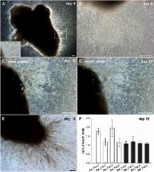
          Results: Vascular outgrowth from the embedded tissue into the fibrin matrix was preceded by leukocyte egress from the tissue fragments. Neo-vessels originating from both the embedded sample and from clusters locally formed by emigrated mononuclear cells were consistently associated with CD45 + leukocytes. MSC and PBMC in co-culture formed vasculogenic clusters. Clusters and cells with endothelial phenotype emerging from them, were surrounded by a collagen IV scaffold. No vascular structures were observed in control 3D monocultures of PBMC or MSC. Paracrine signals released by cultured OA tissue fragments corresponded with elevated levels of granulocyte-colony stimulating factor, vascular endothelial growth factor and interleukin-6 secreted by MSC-PBMC co-cultures.

          Conclusion: Our results show that synovial tissue fragments with immune cell infiltrates have the potential to form new vessels in initially avascular 3D fibrin-based matrices. Cross-talk and cluster formation of MSC with immune cells within the 3D fibrin environment through self-organization and secretion of pro-angiogenic paracrine factors can support neo-vessel growth.

          Related collections

          Most cited references40

          • Record: found
          • Abstract: found
          • Article: not found

          VEGF contributes to postnatal neovascularization by mobilizing bone marrow-derived endothelial progenitor cells.

          Vascular endothelial growth factor (VEGF) has been shown to promote neovascularization in animal models and, more recently, in human subjects. This feature has been assumed to result exclusively from its direct effects on fully differentiated endothelial cells, i.e. angiogenesis. Given its regulatory role in both angiogenesis and vasculogenesis during fetal development, we investigated the hypothesis that VEGF may modulate endothelial progenitor cell (EPC) kinetics for postnatal neovascularization. Indeed, we observed an increase in circulating EPCs following VEGF administration in vivo. VEGF-induced mobilization of bone marrow-derived EPCs resulted in increased differentiated EPCs in vitro and augmented corneal neovascularization in vivo. These findings thus establish a novel role for VEGF in postnatal neovascularization which complements its known impact on angiogenesis.
            Bookmark
            • Record: found
            • Abstract: found
            • Article: not found

            Characterization of two types of endothelial progenitor cells and their different contributions to neovasculogenesis.

            Endothelial progenitor cells (EPC) in one study group is not the same as EPC in other investigators, suggesting that EPC is not a single type of cell population. In this study, we tried to demonstrate the heterogeneity of EPC. We cultured total mononuclear cells from human peripheral blood to get two types of EPC sequentially from the same donors. We called them early EPC and late EPC. Early EPC with spindle shape showed peak growth at 2 to 3 weeks and died at 4 weeks, whereas late EPC with cobblestone shape appeared late at 2 to 3 weeks, showed exponential growth at 4 to 8 weeks, and lived up to 12 weeks. Late EPC was different from early EPC in the expression of VE-cadherin, Flt-1, KDR, and CD45. Late EPC produced more nitric oxide, incorporated more readily into human umbilical vein endothelial cells monolayer, and formed capillary tube better than early EPC. Early EPC secreted angiogenic cytokines (vascular endothelial growth factor, interleukin 8) more so than late EPC during culture in vitro. Both types of EPC showed comparable in vivo vasculogenic capacity. We found two types of EPC from a source of adult peripheral blood that might have different roles in neovasculogenesis based on the identified differences.
              Bookmark
              • Record: found
              • Abstract: found
              • Article: not found

              Pericyte recruitment during vasculogenic tube assembly stimulates endothelial basement membrane matrix formation.

              We show that endothelial cell (EC)-generated vascular guidance tunnels (ie, matrix spaces created during tube formation) serve as conduits for the recruitment and motility of pericytes along EC ablumenal surfaces to facilitate vessel maturation events, including vascular basement membrane matrix assembly and restriction of EC tube diameter. During quail development, pericyte recruitment along microvascular tubes directly correlates with vascular basement membrane matrix deposition. Pericyte recruitment to EC tubes leads to specific induction of fibronectin and nidogen-1 (ie, matrix-bridging proteins that link together basement membrane components) as well as perlecan and laminin isoforms. Coincident with these events, up-regulation of integrins, alpha(5)beta(1), alpha(3)beta(1), alpha(6)beta(1), and alpha(1)beta(1), which bind fibronectin, nidogens, laminin isoforms, and collagen type IV, occurs in EC-pericyte cocultures, but not EC-only cultures. Integrin-blocking antibodies to these receptors, disruption of fibronectin matrix assembly, and small interfering RNA suppression of pericyte tissue inhibitor of metalloproteinase (TIMP)-3 (a known regulator of vascular tube stabilization) all lead to decreased EC basement membrane, resulting in increased vessel lumen diameter, a key indicator of dysfunctional EC-pericyte interactions. Thus, pericyte recruitment to EC-lined tubes during vasculogenesis is a stimulatory event controlling vascular basement membrane matrix assembly, a fundamental maturation step regulating the transition from vascular morphogenesis to stabilization.
                Bookmark

                Author and article information

                Contributors
                Journal
                Front Physiol
                Front Physiol
                Front. Physiol.
                Frontiers in Physiology
                Frontiers Media S.A.
                1664-042X
                05 June 2018
                2018
                : 9
                : 679
                Affiliations
                [1] 1Department of Blood Group Serology and Transfusion Medicine, Medical University of Vienna , Vienna, Austria
                [2] 2Turku Centre for Biotechnology, University of Turku and Åbo Akademi University , Turku, Finland
                [3] 3Department of Orthopedics, Medical University of Vienna , Vienna, Austria
                [4] 4Department of Health Sciences and Biomedicine, Danube University Krems , Krems an der Donau, Austria
                [5] 5Department of Vascular Biology and Thrombosis Research, Center of Physiology and Pharmacology, Medical University of Vienna , Vienna, Austria
                Author notes

                Edited by: Cesare Gargioli, Università degli Studi di Roma Tor Vergata, Italy

                Reviewed by: Stefano Maria Cannata, Università degli Studi di Roma Tor Vergata, Italy; Peggy Lafuste, Université Paris-Est Créteil Val de Marne, France

                *Correspondence: Beate M. Rüger beate.rueger@ 123456meduniwien.ac.at

                This article was submitted to Integrative Physiology, a section of the journal Frontiers in Physiology

                Article
                10.3389/fphys.2018.00679
                5996074
                29922175
                768e6247-d746-466e-9790-50ff50f594b5
                Copyright © 2018 Rüger, Buchacher, Giurea, Kubista, Fischer and Breuss.

                This is an open-access article distributed under the terms of the Creative Commons Attribution License (CC BY). The use, distribution or reproduction in other forums is permitted, provided the original author(s) and the copyright owner are credited and that the original publication in this journal is cited, in accordance with accepted academic practice. No use, distribution or reproduction is permitted which does not comply with these terms.

                History
                : 28 February 2018
                : 15 May 2018
                Page count
                Figures: 8, Tables: 0, Equations: 0, References: 51, Pages: 17, Words: 11042
                Categories
                Physiology
                Original Research

                Anatomy & Physiology
                neovascularization,inflammation,3d fibrin matrix,synovial tissue,mesenchymal stromal cells,self-organization

                Comments

                Comment on this article