1
views
0
recommends
+1 Recommend
0 collections
    0
    shares
      • Record: found
      • Abstract: found
      • Article: not found

      Segmentation of anatomical layers and imaging artifacts in intravascular polarization sensitive optical coherence tomography using attending physician and boundary cardinality losses

      research-article

      Read this article at

      ScienceOpenPublisherPMC
      Bookmark
          There is no author summary for this article yet. Authors can add summaries to their articles on ScienceOpen to make them more accessible to a non-specialist audience.

          Abstract

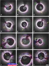
          Intravascular ultrasound and optical coherence tomography are widely available for assessing coronary stenoses and provide critical information to optimize percutaneous coronary intervention. Intravascular polarization-sensitive optical coherence tomography (PS-OCT) measures the polarization state of the light scattered by the vessel wall in addition to conventional cross-sectional images of subsurface microstructure. This affords reconstruction of tissue polarization properties and reveals improved contrast between the layers of the vessel wall along with insight into collagen and smooth muscle content. Here, we propose a convolutional neural network model, optimized using two new loss terms (Boundary Cardinality and Attending Physician), that takes advantage of the additional polarization contrast and classifies the lumen, intima, and media layers in addition to guidewire and plaque shadows. Our model segments the media boundaries through fibrotic plaques and continues to estimate the outer media boundary behind shadows of lipid-rich plaques. We demonstrate that our multi-class classification model outperforms existing methods that exclusively use conventional OCT data, predominantly segment the lumen, and consider subsurface layers at most in regions of minimal disease. Segmentation of all anatomical layers throughout diseased vessels may facilitate stent sizing and will enable automated characterization of plaque polarization properties for investigation of the natural history and significance of coronary atheromas.

          Related collections

          Most cited references63

          • Record: found
          • Abstract: found
          • Article: not found

          A survey on deep learning in medical image analysis

          Deep learning algorithms, in particular convolutional networks, have rapidly become a methodology of choice for analyzing medical images. This paper reviews the major deep learning concepts pertinent to medical image analysis and summarizes over 300 contributions to the field, most of which appeared in the last year. We survey the use of deep learning for image classification, object detection, segmentation, registration, and other tasks. Concise overviews are provided of studies per application area: neuro, retinal, pulmonary, digital pathology, breast, cardiac, abdominal, musculoskeletal. We end with a summary of the current state-of-the-art, a critical discussion of open challenges and directions for future research.
            Bookmark
            • Record: found
            • Abstract: found
            • Article: not found

            Lessons From Sudden Coronary Death

            Arteriosclerosis, Thrombosis, and Vascular Biology, 20(5), 1262-1275
              Bookmark
              • Record: found
              • Abstract: found
              • Article: not found

              U-Net: Convolutional Networks for Biomedical Image Segmentation

              There is large consent that successful training of deep networks requires many thousand annotated training samples. In this paper, we present a network and training strategy that relies on the strong use of data augmentation to use the available annotated samples more efficiently. The architecture consists of a contracting path to capture context and a symmetric expanding path that enables precise localization. We show that such a network can be trained end-to-end from very few images and outperforms the prior best method (a sliding-window convolutional network) on the ISBI challenge for segmentation of neuronal structures in electron microscopic stacks. Using the same network trained on transmitted light microscopy images (phase contrast and DIC) we won the ISBI cell tracking challenge 2015 in these categories by a large margin. Moreover, the network is fast. Segmentation of a 512x512 image takes less than a second on a recent GPU. The full implementation (based on Caffe) and the trained networks are available at http://lmb.informatik.uni-freiburg.de/people/ronneber/u-net . conditionally accepted at MICCAI 2015
                Bookmark

                Author and article information

                Journal
                Biomed Opt Express
                Biomed Opt Express
                BOE
                Biomedical Optics Express
                Optica Publishing Group
                2156-7085
                16 February 2024
                01 March 2024
                : 15
                : 3
                : 1719-1738
                Affiliations
                [1 ]Computer Science and Artificial Intelligence Laboratory (CSAIL), Massachusetts Institute of Technology , Cambridge, MA 02142, USA
                [2 ]Wellman Center for Photomedicine, Massachusetts General Hospital, Harvard Medical School , Boston, MA 02114, USA
                [3 ]Department of Cardiology, Erasmus University Medical Center , Rotterdam, The Netherlands
                [4 ]Division of Cardiovascular Medicine, Department of Medicine, Brigham and Women’s Hospital, Harvard Medical School , Boston, MA 02115, USA
                [5 ]Institute for Medical Engineering and Science, Massachusetts Institute of Technology , Cambridge, MA 02142, USA
                Author notes
                [†]

                The authors contributed equally to this work.

                Author information
                https://orcid.org/0000-0002-8551-5008
                https://orcid.org/0000-0003-3819-1271
                Article
                514673
                10.1364/BOE.514673
                10942710
                38495711
                73421c62-ee7e-44eb-beb5-e4de2b2d72d2
                © 2024 Optica Publishing Group

                https://doi.org/10.1364/OA_License_v2#VOR-OA

                © 2024 Optica Publishing Group under the terms of the Optica Open Access Publishing Agreement

                History
                : 29 November 2023
                : 03 February 2024
                : 04 February 2024
                Funding
                Funded by: Simard Fund
                Funded by: Robert M. McCormick Tribune Foundation 10.13039/100006359
                Award ID: Charitable Fund
                Funded by: Massachusetts General Hospital 10.13039/100005294
                Award ID: Bullock Postdoctoral Fellowship
                Funded by: American Heart Association 10.13039/100000968
                Award ID: 18CSA34080399
                Funded by: National Institutes of Health 10.13039/100000002
                Award ID: 1R01HL134892
                Award ID: 1R01HL163099-01
                Award ID: P41EB-015902
                Award ID: P41EB-015903
                Categories
                Article

                Vision sciences
                Vision sciences

                Comments

                Comment on this article