0
views
0
recommends
+1 Recommend
0 collections
    0
    shares
      • Record: found
      • Abstract: found
      • Article: found
      Is Open Access

      Disruption of the mitochondrial network in a mouse model of Huntington's disease visualized by in-tissue multiscale 3D electron microscopy

      research-article

      Read this article at

      Bookmark
          There is no author summary for this article yet. Authors can add summaries to their articles on ScienceOpen to make them more accessible to a non-specialist audience.

          Abstract

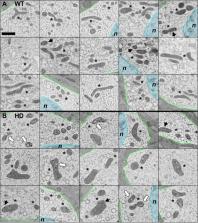
          Huntington’s disease (HD) is an inherited neurodegenerative disorder caused by an expanded CAG repeat in the coding sequence of huntingtin protein. Initially, it predominantly affects medium-sized spiny neurons (MSSNs) of the corpus striatum. No effective treatment is still available, thus urging the identification of potential therapeutic targets. While evidence of mitochondrial structural alterations in HD exists, previous studies mainly employed 2D approaches and were performed outside the strictly native brain context. In this study, we adopted a novel multiscale approach to conduct a comprehensive 3D in situ structural analysis of mitochondrial disturbances in a mouse model of HD. We investigated MSSNs within brain tissue under optimal structural conditions utilizing state-of-the-art 3D imaging technologies, specifically FIB/SEM for the complete imaging of neuronal somas and Electron Tomography for detailed morphological examination, and image processing-based quantitative analysis. Our findings suggest a disruption of the mitochondrial network towards fragmentation in HD. The network of interlaced, slim and long mitochondria observed in healthy conditions transforms into isolated, swollen and short entities, with internal cristae disorganization, cavities and abnormally large matrix granules.

          Supplementary Information

          The online version contains supplementary material available at 10.1186/s40478-024-01802-2.

          Related collections

          Most cited references66

          • Record: found
          • Abstract: found
          • Article: found
          Is Open Access

          seaborn: statistical data visualization

            Bookmark
            • Record: found
            • Abstract: found
            • Article: not found

            Computer visualization of three-dimensional image data using IMOD.

            We have developed a computer software package, IMOD, as a tool for analyzing and viewing three-dimensional biological image data. IMOD is useful for studying and modeling data from tomographic, serial section, and optical section reconstructions. The software allows image data to be visualized by several different methods. Models of the image data can be visualized by volume or contour surface rendering and can yield quantitative information.
              Bookmark
              • Record: found
              • Abstract: not found
              • Article: not found

              A novel gene containing a trinucleotide repeat that is expanded and unstable on Huntington's disease chromosomes

                Bookmark

                Author and article information

                Contributors
                MR.Fernandez@csic.es
                JJ.Fernandez@csic.es
                Journal
                Acta Neuropathol Commun
                Acta Neuropathol Commun
                Acta Neuropathologica Communications
                BioMed Central (London )
                2051-5960
                5 June 2024
                5 June 2024
                2024
                : 12
                : 88
                Affiliations
                [1 ]Department of Psychiatry, University of Pittsburgh, ( https://ror.org/01an3r305) Pittsburgh, PA 15213 USA
                [2 ]Advanced Microscopy Laboratory, University of Zaragoza, ( https://ror.org/012a91z28) Zaragoza, Spain
                [3 ]GRID grid.11205.37, ISNI 0000 0001 2152 8769, Instituto de Nanociencia y Materiales de Aragon (INMA), , CSIC-Universidad de Zaragoza, ; 50018 Zaragoza, Spain
                [4 ]Department of Condensed Matter Physics, University of Zaragoza, ( https://ror.org/012a91z28) Zaragoza, Spain
                [5 ]Department of Chemistry and Chemical Biology, Rutgers, The State University of New Jersey, ( https://ror.org/05vt9qd57) Piscataway, NJ 08854 USA
                [6 ]Spanish National Research Council (CSIC, CINN), Health Research Institute of Asturias (ISPA), ( https://ror.org/02gfc7t72) 33011 Oviedo, Spain
                Author information
                http://orcid.org/0000-0003-2222-3355
                Article
                1802
                10.1186/s40478-024-01802-2
                11151585
                38840253
                72d84c64-9b2d-4ae7-9989-35cb5f71f6b1
                © The Author(s) 2024

                Open Access This article is licensed under a Creative Commons Attribution 4.0 International License, which permits use, sharing, adaptation, distribution and reproduction in any medium or format, as long as you give appropriate credit to the original author(s) and the source, provide a link to the Creative Commons licence, and indicate if changes were made. The images or other third party material in this article are included in the article's Creative Commons licence, unless indicated otherwise in a credit line to the material. If material is not included in the article's Creative Commons licence and your intended use is not permitted by statutory regulation or exceeds the permitted use, you will need to obtain permission directly from the copyright holder. To view a copy of this licence, visit http://creativecommons.org/licenses/by/4.0/. The Creative Commons Public Domain Dedication waiver ( http://creativecommons.org/publicdomain/zero/1.0/) applies to the data made available in this article, unless otherwise stated in a credit line to the data.

                History
                : 4 April 2024
                : 27 May 2024
                Funding
                Funded by: FundRef http://dx.doi.org/10.13039/501100011033, Agencia Estatal de Investigación;
                Award ID: PID2022-139071NB-I00
                Award Recipient :
                Funded by: Consejo Superior de Investigaciones Cientificas (CSIC)
                Categories
                Research
                Custom metadata
                © BioMed Central Ltd., part of Springer Nature 2024

                3d electron microscopy,volume electron microscopy,electron tomography,cryo-fixation,huntington's disease,mitochondria

                Comments

                Comment on this article