23
views
0
recommends
+1 Recommend
0 collections
    0
    shares
      • Record: found
      • Abstract: found
      • Article: found
      Is Open Access

      Yellow nail syndrome: a review

      review-article

      Read this article at

      Bookmark
          There is no author summary for this article yet. Authors can add summaries to their articles on ScienceOpen to make them more accessible to a non-specialist audience.

          Abstract

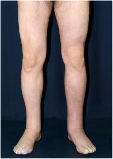
          Yellow nail syndrome (YNS; OMIM 153300, ORPHA662) is a very rare disorder that almost always occurs after 50 years of age but a juvenile or familial form has also been observed. YNS is diagnosed based on a triad associating yellow nail discoloration, pulmonary manifestations (chronic cough, bronchiectasia, pleural effusion) and lower limb lymphedema. Chronic sinusitis is frequently associated with the triad. YNS etiology remains unknown but a role of lymphatic impairment is usually evoked. YNS is more frequently isolated but may be associated in rare cases with autoimmune diseases, other clinical manifestations implicating lymphatic functions or cancer and, hence, is also considered a paraneoplastic syndrome. YNS management is symptomatic and not codified. YNS can resolve spontaneously. Oral vitamin E alone or even better when associated with triazole antifungals may achieve partial or total disappearance of nail discoloration. Pleural effusion can be treated surgically, with decortication/pleurectomy or pleurodesis. Antibiotic prophylaxis is prescribed for bronchiectasia with chronic sputum production. Lymphedema treatment is based on low-stretch bandages and the wearing of elastic compression garments combined with skin care, exercises and, as needed, manual lymph drainage.

          Related collections

          Most cited references132

          • Record: found
          • Abstract: found
          • Article: not found

          Risk factors for erysipelas of the leg (cellulitis): case-control study.

          To assess risk factors for erysipelas of the leg (cellulitis). Case-control study. 7 hospital centres in France. 167 patients admitted to hospital for erysipelas of the leg and 294 controls. In multivariate analysis, a disruption of the cutaneous barrier (leg ulcer, wound, fissurated toe-web intertrigo, pressure ulcer, or leg dermatosis) (odds ratio 23.8, 95% confidence interval 10.7 to 52.5), lymphoedema (71.2, 5.6 to 908), venous insufficiency (2.9, 1.0 to 8.7), leg oedema (2.5, 1.2 to 5.1) and being overweight (2.0, 1.1 to 3.7) were independently associated with erysipelas of the leg. No association was observed with diabetes, alcohol, or smoking. Population attributable risk for toe-web intertrigo was 61%. This first case-control study highlights the major role of local risk factors (mainly lymphoedema and site of entry) in erysipelas of the leg. From a public health perspective, detecting and treating toe-web intertrigo should be evaluated in the secondary prevention of erysipelas of the leg.
            Bookmark
            • Record: found
            • Abstract: found
            • Article: not found

            A systematic review of the evidence for complete decongestive therapy in the treatment of lymphedema from 2004 to 2011.

            To critically analyze the contemporary published research that pertains to the individual components of complete decongestive therapy (CDT), as well as CDT as a bundled intervention in the treatment of lymphedema. Publications were retrieved from 11 major medical indices for articles published from 2004-2010 by using search terms for lymphedema and management approaches. Literature archives of the authors and reference lists were examined through 2011. A research librarian assisted with initial literature searches by using search terms used in the Best Practice for the Management of Lymphoedema, plus expanded terms, for literature related to lymphedema. Authors sorted relevant literature for inclusion and exclusion; included articles were sorted into topical areas for data extraction and assessment of level of evidence by using a published grading system and consensus process. The authors reviewed 99 articles, of which 26 met inclusion criteria for individual studies and 1 case study did not meet strict inclusion criteria. In addition, 14 review articles and 2 consensus articles were reviewed. Information on study design and/or objectives, participants, outcomes, intervention, results, and study strengths and weaknesses was extracted from each article. Study evidence was categorized according to the Oncology Nursing Society Putting Evidence into Practice level of evidence guidelines after achieving consensus among authors. Levels of evidence were only moderately strong, because there were few randomized controlled trials with control groups, well-controlled interventions, and precise measurements of volume, mobility and/or function, and quality of life. Treatment interventions were often bundled, which makes it difficult to determine the contribution of each individual component of treatment to the outcomes achieved. CDT is seen to be effective in reducing lymphedema. This review focuses on original research about CDT as a bundled intervention and 2 individual components, manual lymph drainage and compression bandages. Additional studies are needed to determine the value and efficacy of the other individual components of CDT. Copyright © 2012 American Academy of Physical Medicine and Rehabilitation. Published by Elsevier Inc. All rights reserved.
              Bookmark
              • Record: found
              • Abstract: found
              • Article: not found

              Regulation of adipogenesis by lymphatic fluid stasis: part I. Adipogenesis, fibrosis, and inflammation.

              Although fat deposition is a defining clinical characteristic of lymphedema, the cellular mechanisms that regulate this response remain unknown. The goals of this two-part study were to determine the effect of lymphatic fluid stasis on adipogenesis and inflammation (part I) and how these changes regulate the temporal and spatial expression of fat differentiation genes (part II). Adult female mice underwent tail lymphatic ablation and were euthanized 6 weeks after surgery (n = 20). Fat deposition, fibrosis, and inflammation were then analyzed in the regions of the tail exposed to lymphatic fluid stasis as compared with normal lymphatic flow. Lymphatic fluid stasis in the tail resulted in significant subcutaneous fat deposition, with a 2-fold increase in fat thickness (p < 0.01). In addition, lymphatic stasis was associated with subcutaneous fat fibrosis and collagen deposition. Adipogenesis in response to lymphatic fluid stasis was associated with a marked mononuclear cell inflammatory response (5-fold increase in CD45 cells; p < 0.001). In addition, the authors noted a significant increase in the number of monocytes/macrophages as identified by F4/80 immunohistochemistry (p < 0.001). The mouse-tail model has pathologic findings that are similar to clinical lymphedema, including fat deposition, fibrosis, and inflammation. Adipogenesis in response to lymphatic fluid stasis closely resembles this process in obesity. This model therefore provides an excellent means with which to study the molecular mechanisms that regulate the pathophysiology of lymphedema.
                Bookmark

                Author and article information

                Contributors
                +33 (0) 145 30 82 35 , stephane.vignes@cognacq-jay.fr
                Journal
                Orphanet J Rare Dis
                Orphanet J Rare Dis
                Orphanet Journal of Rare Diseases
                BioMed Central (London )
                1750-1172
                27 February 2017
                27 February 2017
                2017
                : 12
                : 42
                Affiliations
                [1 ]Department of Lymphology, Centre National de Référence des Maladies Vasculaires Rares (Lymphœdèmes primaires), Hôpital Cognacq-Jay, 15, rue Eugène-Millon, 75015 Paris, France
                [2 ]ISNI 0000 0001 2188 3779, GRID grid.7459.f, , Nail Disease Centre, ; 42, rue des Serbes, 06400 Cannes, France
                Article
                594
                10.1186/s13023-017-0594-4
                5327582
                28241848
                716c9384-71ef-4737-ac0b-4df80251c513
                © The Author(s). 2017

                Open AccessThis article is distributed under the terms of the Creative Commons Attribution 4.0 International License ( http://creativecommons.org/licenses/by/4.0/), which permits unrestricted use, distribution, and reproduction in any medium, provided you give appropriate credit to the original author(s) and the source, provide a link to the Creative Commons license, and indicate if changes were made. The Creative Commons Public Domain Dedication waiver ( http://creativecommons.org/publicdomain/zero/1.0/) applies to the data made available in this article, unless otherwise stated.

                History
                : 26 October 2016
                : 21 February 2017
                Categories
                Review
                Custom metadata
                © The Author(s) 2017

                Infectious disease & Microbiology
                yellow nail syndrome,respiratory manifestations,sinusitis,lymphedema,review

                Comments

                Comment on this article