16
views
0
recommends
+1 Recommend
0 collections
    0
    shares
      • Record: found
      • Abstract: found
      • Article: found
      Is Open Access

      Ultrasonography of the Kidneys in Healthy and Diseased Camels (Camelus dromedarius)

      review-article
      1 , 2 ,
      Veterinary Medicine International
      Hindawi

      Read this article at

      Bookmark
          There is no author summary for this article yet. Authors can add summaries to their articles on ScienceOpen to make them more accessible to a non-specialist audience.

          Abstract

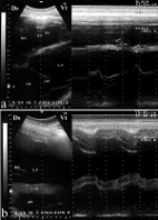
          This review article is written to describe the results of ultrasonography of the kidneys in healthy camels as well as camels with some renal disorders. In the dromedary camel, the physiology of the kidney is of interest in view of the specialization of the camel to hot dry deserts and to prolonged periods without water. It plays an important role in water conservation through the production of highly concentrated urine that may predispose animal to varieties of renal disorders. Examples of kidney affections in dromedary camels are renal capsular pigmentation, medullary hyperemia, subcapsular calcification, cortical and medullar discoloration, hemorrhage in renal pelvis, nephrolithiasis, and hydatidosis. Congestion, hemorrhage, hydronephrosis, acute glomerulonephritis, subacute glomerulonephritis, chronic glomerulonephritis, diffuse interstitial nephritis, focal interstitial nephritis, renal cyst, hyaline degeneration, renal amyloidosis, tubular nephrosis, pyelonephritis, hemosiderosis, and renal toxicity. When the kidney is examined by ultrasonography, the clinician can get sufficient information about the size, position, and echo patterns of the renal cortex and medulla and renal pelvis and outlines of the renal blood vessels. In recent years, ultrasonography has been used in camels for scanning of the healthy status as well as evaluation and determining the diagnosis and prognosis of diseased cases. Examples of diseases evaluated by ultrasonography are paratuberculosis, trypanosomiasis, pneumonia, pleurisy, gastrointestinal neoplasms, chronic peritonitis, splenic abscessation, and hepatic disorders. Of the renal disorders assessed by ultrasonography are nephrolithiasis, hydronephrosis, pyelonephritis, renal abscessation, and renal neoplasms. Ultrasound guidance in biopsy of renal specimens has also been reported in dromedary camels.

          Related collections

          Most cited references47

          • Record: found
          • Abstract: found
          • Article: not found

          A herd level analysis of urinary tract infection in dairy cattle.

          Cystitis, urethritis and pyelonephritis in cattle most commonly result from ascending urinary tract infection with Corynebacterium renale, Corynebacterium cystidis, Corynebacterium pilosum or Escherichia coli. We describe the clinical, bacteriological, clinical-pathological and epidemiological findings in a dairy cattle herd with urinary tract infection (UTI). Blood and urine samples from 17 calves and 19 cows were submitted to laboratory examinations. Depression, muscle wasting, weakness and frequent urine dribbling were the main characteristics of UTI in calves. Affected cows showed weight loss and an abrupt reduction in feed intake and milk production. Enlargement of the left kidney and loss of normal lobulation were evident on rectal examination. E. coli was the most frequent cause of UTI but C. renale, alpha-haemolytic Streptococcus spp., Proteus spp. Pseudomonas aeruginosa, Klebsiella spp. and Oligella urethralis were isolated as well. Differences in total protein and several protein fractions were found between affected and healthy animals.
            Bookmark
            • Record: found
            • Abstract: found
            • Article: found
            Is Open Access

            Echocardiography of the normal camel (Camelus dromedaries) heart: technique and cardiac dimensions

            Background Echocardiography and intra-cardiac dimensions have not previously been reported in adult camels despite its potential application for medical purpose. The aim of this study was to describe the results of a prospective study, aiming to report normal cardiac appearance and normal chamber dimensions in adult camels (Camelus dromedarius). Results On the right side, when the probe was placed in the 5th or 4th intercostal space (ICS), the caudal long-axis four-chamber view of the ventricles, atria, and the interventricular septum was obtained. Placing the probe slightly more cranially in the 4th ICS, the caudal long-axis four-chamber view and the caudal long-axis view of the left ventricular outflow tract (LVOT) were imaged. In 7 camels, a hybrid view between a “four-chamber” and “LVOT view” was imaged from the same position. The short-axis view of the ventricles was obtained in the 4th ICS where the transducer was rotated between 0° and 25°. Placement of the transducer in the 3rd ICS allowed visualisation of the right ventricular outflow tract (RVOT). On the left side, when the probe was placed in the 5th or 4th ICS, a four-chamber view was obtained. The LVOT is imaged in the 4th ICS and the RVOT was seen from the 3rd ICS. Conclusions This study showed that it is possible to obtain good-quality echocardiograms in adult camels and provide normal cardiac dimensions. This study could be used as a reference for further studies concerning camels with cardiac diseases.
              Bookmark
              • Record: found
              • Abstract: found
              • Article: not found

              Clinical, ultrasonographic, and pathologic findings in 70 camels (Camelus dromedarius) with Johne's disease.

              This study evaluated the use of ultrasonography for the diagnosis of Johne's disease in camels (Camelus dromedarius). Seventy camels with confirmed Johne's disease were examined by ultrasonography and subsequent necropsy; 15 healthy camels were included as controls. The most outstanding findings were visible enlargement of the mesenteric lymph nodes in 52 (74%) camels. Lesions had either echogenic (26%; n = 18) or anechoic (69%; n = 48) capsule and the contents were either anechoic (21%; n = 15), echogenic (27%; n = 19), or heterogeneous (46%; n = 32). Clumps of echogenic tissue interspersed with fluid pockets were imaged between the intestinal loops in 9 (13%) camels. There was mild, moderate, or severe thickening and corrugation of the intestinal wall, excessive anechoic fluid in the abdominal cavity in 18 (26%) camels, increased hepatic brightness in 30 (43%) camels, and pericardial and pleural effusions in 22 (31%) camels. Sensitivity values for detecting intestinal lesions and enlarged mesenteric lymph nodes were 95% and 84%, respectively.
                Bookmark

                Author and article information

                Contributors
                Journal
                Vet Med Int
                Vet Med Int
                VMI
                Veterinary Medicine International
                Hindawi
                2090-8113
                2042-0048
                2020
                21 October 2020
                : 2020
                : 7814927
                Affiliations
                1Department of Veterinary Medicine, College of Agriculture and Veterinary Medicine, Qassim University, P.O. Box 6622, Buraidah, 51452, Saudi Arabia
                2Department of Animal Medicine, Faculty of Veterinary Medicine, Zagazig University, 44519, Zagazig, Egypt
                Author notes

                Academic Editor: Antonio Ortega-Pacheco

                Author information
                https://orcid.org/0000-0002-3796-9590
                Article
                10.1155/2020/7814927
                7596541
                33144934
                71292271-861f-4285-9a33-cc8054be0659
                Copyright © 2020 Mohamed Tharwat.

                This is an open access article distributed under the Creative Commons Attribution License, which permits unrestricted use, distribution, and reproduction in any medium, provided the original work is properly cited.

                History
                : 18 March 2020
                : 15 September 2020
                : 17 September 2020
                Funding
                Funded by: Qassim University
                Categories
                Review Article

                Veterinary medicine
                Veterinary medicine

                Comments

                Comment on this article