21
views
0
recommends
+1 Recommend
0 collections
    0
    shares
      • Record: found
      • Abstract: found
      • Article: found
      Is Open Access

      3D Differentiation of Neural Stem Cells in Macroporous Photopolymerizable Hydrogel Scaffolds

      research-article
      , , *
      PLoS ONE
      Public Library of Science

      Read this article at

      Bookmark
          There is no author summary for this article yet. Authors can add summaries to their articles on ScienceOpen to make them more accessible to a non-specialist audience.

          Abstract

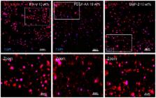
          Neural stem/progenitor cells (NSPCs) are the stem cell of the adult central nervous system (CNS). These cells are able to differentiate into the major cell types found in the CNS (neurons, oligodendrocytes, astrocytes), thus NSPCs are the mechanism by which the adult CNS could potentially regenerate after injury or disorder. Microenviromental factors are critical for guiding NSPC differentiation and are thus important for neural tissue engineering. In this study, D-mannitol crystals were mixed with photocrosslinkable methacrylamide chitosan (MAC) as a porogen to enhance pore size during hydrogel formation. D-mannitol was admixed to MAC at 5, 10 and 20 wt% D-mannitol per total initial hydrogel weight. D-mannitol crystals were observed to dissolve and leave the scaffold within 1 hr. Quantification of resulting average pore sizes showed that D-mannitol addition resulted in larger average pore size (5 wt%, 4060±160 µm 2, 10 wt%, 6330±1160 µm 2, 20 wt%, 7600±1550 µm 2) compared with controls (0 wt%, 3150±220 µm 2). Oxygen diffusion studies demonstrated that larger average pore area resulted in enhanced oxygen diffusion through scaffolds. Finally, the differentiation responses of NSPCs to phenotypic differentiation conditions were studied for neurons, astrocytes and oligodendrocytes in hydrogels of varied porosity over 14 d. Quantification of total cell numbers at day 7 and 14, showed that cell numbers decreased with increased porosity and over the length of the culture. At day 14 immunohistochemistry quantification for primary cell types demonstrated significant differentiation to the desired cells types, and that total percentages of each cell type was greatest when scaffolds were more porous. These results suggest that larger pore sizes in MAC hydrogels effectively promote NSPC 3D differentiation.

          Related collections

          Most cited references52

          • Record: found
          • Abstract: found
          • Article: not found

          Generation of neurons and astrocytes from isolated cells of the adult mammalian central nervous system.

          Neurogenesis in the mammalian central nervous system is believed to end in the period just after birth; in the mouse striatum no new neurons are produced after the first few days after birth. In this study, cells isolated from the striatum of the adult mouse brain were induced to proliferate in vitro by epidermal growth factor. The proliferating cells initially expressed nestin, an intermediate filament found in neuroepithelial stem cells, and subsequently developed the morphology and antigenic properties of neurons and astrocytes. Newly generated cells with neuronal morphology were immunoreactive for gamma-aminobutyric acid and substance P, two neurotransmitters of the adult striatum in vivo. Thus, cells of the adult mouse striatum have the capacity to divide and differentiate into neurons and astrocytes.
            Bookmark
            • Record: found
            • Abstract: found
            • Article: not found

            Biology of oligodendrocyte and myelin in the mammalian central nervous system.

            Oligodendrocytes, the myelin-forming cells of the central nervous system (CNS), and astrocytes constitute macroglia. This review deals with the recent progress related to the origin and differentiation of the oligodendrocytes, their relationships to other neural cells, and functional neuroglial interactions under physiological conditions and in demyelinating diseases. One of the problems in studies of the CNS is to find components, i.e., markers, for the identification of the different cells, in intact tissues or cultures. In recent years, specific biochemical, immunological, and molecular markers have been identified. Many components specific to differentiating oligodendrocytes and to myelin are now available to aid their study. Transgenic mice and spontaneous mutants have led to a better understanding of the targets of specific dys- or demyelinating diseases. The best examples are the studies concerning the effects of the mutations affecting the most abundant protein in the central nervous myelin, the proteolipid protein, which lead to dysmyelinating diseases in animals and human (jimpy mutation and Pelizaeus-Merzbacher disease or spastic paraplegia, respectively). Oligodendrocytes, as astrocytes, are able to respond to changes in the cellular and extracellular environment, possibly in relation to a glial network. There is also a remarkable plasticity of the oligodendrocyte lineage, even in the adult with a certain potentiality for myelin repair after experimental demyelination or human diseases.
              Bookmark
              • Record: found
              • Abstract: found
              • Article: not found

              Porous chitosan scaffolds for tissue engineering.

              The wide array of tissue engineering applications exacerbates the need for biodegradable materials with broad potential. Chitosan, the partially deacetylated derivative of chitin, may be one such material. In this study, we examined the use of chitosan for formation of porous scaffolds of controlled microstructure in several tissue-relevant geometries. Porous chitosan materials were prepared by controlled freezing and lyophilization of chitosan solutions and gels. The materials were characterized via light and scanning electron microscopy as well as tensile testing. The scaffolds formed included porous membranes, blocks, tubes and beads. Mean pore diameters could be controlled within the range 1-250 microm, by varying the freezing conditions. Freshly lyophilized chitosan scaffolds could be treated with glycosaminoglycans to form ionic complex materials which retained the original pore structure. Chitosan scaffolds could be rehydrated via an ethanol series to avoid the stiffening caused by rehydration in basic solutions. Hydrated porous chitosan membranes were at least twice as extensible as non-porous chitosan membranes, but their elastic moduli and tensile strengths were about tenfold lower than non-porous controls. The methods and structures described here provide a starting point for the design and fabrication of a family of polysaccharide based scaffold materials with potentially broad applicability.
                Bookmark

                Author and article information

                Contributors
                Role: Editor
                Journal
                PLoS One
                PLoS ONE
                plos
                plosone
                PLoS ONE
                Public Library of Science (San Francisco, USA )
                1932-6203
                2012
                7 November 2012
                : 7
                : 11
                : e48824
                Affiliations
                [1]Department of Chemical and Biomolecular Engineering, The University of Akron, Akron, Ohio, United States of America
                University of Milan-Bicocca, Italy
                Author notes

                Competing Interests: The authors have read the journal's policy and have the following conflicts: Dr. Nic D. Leipzig is a PLOS ONE Editorial Board member. This does not alter the authors' adherence to all the PLOS ONE policies on sharing data and materials.

                Conceived and designed the experiments: HL AW NDL. Performed the experiments: HL AW. Analyzed the data: HL AW NDL. Contributed reagents/materials/analysis tools: NDL. Wrote the paper: HL AW NDL.

                Article
                PONE-D-12-08827
                10.1371/journal.pone.0048824
                3492243
                23144988
                700fe40d-8409-4dff-b71b-c3c698e566d1
                Copyright @ 2012

                This is an open-access article distributed under the terms of the Creative Commons Attribution License, which permits unrestricted use, distribution, and reproduction in any medium, provided the original author and source are credited.

                History
                : 26 March 2012
                : 4 October 2012
                Page count
                Pages: 11
                Funding
                The work was supported by University of Akron startup funding. The funders had no role in study design, data collection and analysis, decision to publish, or preparation of the manuscript.
                Categories
                Research Article
                Biology
                Biotechnology
                Bioengineering
                Biomedical Engineering
                Biomaterials
                Tissue Engineering
                Developmental Biology
                Stem Cells
                Adult Stem Cells
                Model Organisms
                Animal Models
                Rat
                Neuroscience
                Developmental Neuroscience
                Neural Stem Cells
                Engineering
                Bioengineering
                Biomedical Engineering
                Materials Science
                Biomaterials

                Uncategorized
                Uncategorized

                Comments

                Comment on this article