15
views
0
recommends
+1 Recommend
0 collections
    0
    shares
      • Record: found
      • Abstract: found
      • Article: found
      Is Open Access

      Mutant p53 partners in crime

      review-article
      1 , 2 , 2 , *
      Cell Death and Differentiation
      Nature Publishing Group

      Read this article at

      Bookmark
          There is no author summary for this article yet. Authors can add summaries to their articles on ScienceOpen to make them more accessible to a non-specialist audience.

          Abstract

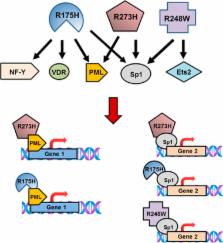
          Mutant p53 proteins impart changes in cellular behavior and function through interactions with proteins that alter gene expression. The milieu of intracellular proteins available to interact with mutant p53 is context specific and changes with disease, cell type, and environmental conditions. Varying conformations of mutant p53 largely dictate protein–protein interactions as different point mutations within protein-coding regions greatly alter the extent and array of gain-of-function (GOF) activities. Given such variables, how can knowledge regarding p53 missense mutations be translated into predicting or altering biologic activity for therapy? How may knowledge regarding mutant p53 functions within certain disease contexts be harnessed to blunt or ablate mutant p53 GOF for therapy? In this article, we review known proteins that interact with mutant p53 and result in the activation of genes that contribute to p53 GOF with particular emphasis on context dependency and an evolving appreciation of GOF mechanisms.

          Related collections

          Most cited references36

          • Record: found
          • Abstract: found
          • Article: not found

          Mutant p53: one name, many proteins.

          There is now strong evidence that mutation not only abrogates p53 tumor-suppressive functions, but in some instances can also endow mutant proteins with novel activities. Such neomorphic p53 proteins are capable of dramatically altering tumor cell behavior, primarily through their interactions with other cellular proteins and regulation of cancer cell transcriptional programs. Different missense mutations in p53 may confer unique activities and thereby offer insight into the mutagenic events that drive tumor progression. Here we review mechanisms by which mutant p53 exerts its cellular effects, with a particular focus on the burgeoning mutant p53 transcriptome, and discuss the biological and clinical consequences of mutant p53 gain of function.
            Bookmark
            • Record: found
            • Abstract: found
            • Article: not found

            Mutant p53 gain of function in two mouse models of Li-Fraumeni syndrome.

            The p53 tumor suppressor gene is commonly altered in human tumors, predominantly through missense mutations that result in accumulation of mutant p53 protein. These mutations may confer dominant-negative or gain-of-function properties to p53. To ascertain the physiological effects of p53 point mutation, the structural mutant p53R172H and the contact mutant p53R270H (codons 175 and 273 in humans) were engineered into the endogenous p53 locus in mice. p53R270H/+ and p53R172H/+ mice are models of Li-Fraumeni Syndrome; they developed allele-specific tumor spectra distinct from p53+/- mice. In addition, p53R270H/- and p53R172H/- mice developed novel tumors compared to p53-/- mice, including a variety of carcinomas and more frequent endothelial tumors. Dominant effects that varied by allele and function were observed in primary cells derived from p53R270H/+ and p53R172H/+ mice. These results demonstrate that point mutant p53 alleles expressed under physiological control have enhanced oncogenic potential beyond the simple loss of p53 function.
              Bookmark
              • Record: found
              • Abstract: found
              • Article: not found

              Gain of function of a p53 hot spot mutation in a mouse model of Li-Fraumeni syndrome.

              Individuals with Li-Fraumeni syndrome carry inherited mutations in the p53 tumor suppressor gene and are predisposed to tumor development. To examine the mechanistic nature of these p53 missense mutations, we generated mice harboring a G-to-A substitution at nucleotide 515 of p53 (p53+/515A) corresponding to the p53R175H hot spot mutation in human cancers. Although p53+/515A mice display a similar tumor spectrum and survival curve as p53+/- mice, tumors from p53+/515A mice metastasized with high frequency. Correspondingly, the embryonic fibroblasts from the p53515A/515A mutant mice displayed enhanced cell proliferation, DNA synthesis, and transformation potential. The disruption of p63 and p73 in p53-/- cells increased transformation capacity and reinitiated DNA synthesis to levels observed in p53515A/515A cells. Additionally, p63 and p73 were functionally inactivated in p53515A cells. These results provide in vivo validation for the gain-of-function properties of certain p53 missense mutations and suggest a mechanistic basis for these phenotypes.
                Bookmark

                Author and article information

                Journal
                Cell Death Differ
                Cell Death Differ
                Cell Death and Differentiation
                Nature Publishing Group
                1350-9047
                1476-5403
                January 2018
                03 November 2017
                1 January 2018
                : 25
                : 1
                : 161-168
                Affiliations
                [1 ]Department of Surgical Oncology, The University of Texas MD Anderson Cancer Center , Houston, TX 77030, USA
                [2 ]Department of Genetics, The University of Texas MD Anderson Cancer Center , Houston, TX 77030, USA
                Author notes
                [* ]Department of Genetics, The University of Texas MD Anderson Cancer Center , Houston, TX 77030, USA. Tel: +1 713 834 6386 Fax: +1 713 834 6380. E-mail: gglozano@ 123456mdanderson.org
                Article
                cdd2017185
                10.1038/cdd.2017.185
                5729539
                29099488
                6ff7b712-c382-4f09-8240-107a6b4c7a97
                Copyright © 2018 The Author(s)

                This work is licensed under a Creative Commons Attribution-NonCommercial-NoDerivs 4.0 International License. The images or other third party material in this article are included in the article’s Creative Commons license, unless indicated otherwise in the credit line; if the material is not included under the Creative Commons license, users will need to obtain permission from the license holder to reproduce the material. To view a copy of this license, visit http://creativecommons.org/licenses/by-nc-nd/4.0/

                History
                : 21 June 2017
                : 15 September 2017
                : 18 September 2017
                Categories
                Review

                Cell biology
                Cell biology

                Comments

                Comment on this article