12
views
0
recommends
+1 Recommend
0 collections
    0
    shares
      • Record: found
      • Abstract: found
      • Article: found
      Is Open Access

      Location-Specific Comparison Between a 3D In-Stent Restenosis Model and Micro-CT and Histology Data from Porcine In Vivo Experiments

      research-article

      Read this article at

      Bookmark
          There is no author summary for this article yet. Authors can add summaries to their articles on ScienceOpen to make them more accessible to a non-specialist audience.

          Abstract

          Background

          Coronary artery restenosis is an important side effect of percutaneous coronary intervention. Computational models can be used to better understand this process. We report on an approach for validation of an in silico 3D model of in-stent restenosis in porcine coronary arteries and illustrate this approach by comparing the modelling results to in vivo data for 14 and 28 days post-stenting.

          Methods

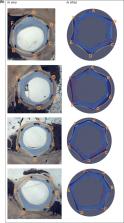
          This multiscale model includes single-scale models for stent deployment, blood flow and tissue growth in the stented vessel, including smooth muscle cell (SMC) proliferation and extracellular matrix (ECM) production. The validation procedure uses data from porcine in vivo experiments, by simulating stent deployment using stent geometry obtained from micro computed tomography (micro-CT) of the stented vessel and directly comparing the simulation results of neointimal growth to histological sections taken at the same locations.

          Results

          Metrics for comparison are per-strut neointimal thickness and per-section neointimal area. The neointimal area predicted by the model demonstrates a good agreement with the detailed experimental data. For 14 days post-stenting the relative neointimal area, averaged over all vessel sections considered, was 20 ± 3% in vivo and 22 ± 4% in silico. For 28 days, the area was 42 ± 3% in vivo and 41 ± 3% in silico.

          Conclusions

          The approach presented here provides a very detailed, location-specific, validation methodology for in silico restenosis models. The model was able to closely match both histology datasets with a single set of parameters. Good agreement was obtained for both the overall amount of neointima produced and the local distribution. It should be noted that including vessel curvature and ECM production in the model was paramount to obtain a good agreement with the experimental data.

          Electronic supplementary material

          The online version of this article (10.1007/s13239-019-00431-4) contains supplementary material, which is available to authorized users.

          Related collections

          Most cited references50

          • Record: found
          • Abstract: found
          • Article: not found

          The role of shear stress in the pathogenesis of atherosclerosis.

          Although the pathobiology of atherosclerosis is a complex multifactorial process, blood flow-induced shear stress has emerged as an essential feature of atherogenesis. This fluid drag force acting on the vessel wall is mechanotransduced into a biochemical signal that results in changes in vascular behavior. Maintenance of a physiologic, laminar shear stress is known to be crucial for normal vascular functioning, which includes the regulation of vascular caliber as well as inhibition of proliferation, thrombosis and inflammation of the vessel wall. Thus, shear stress is atheroprotective. It is also recognized that disturbed or oscillatory flows near arterial bifurcations, branch ostia and curvatures are associated with atheroma formation. Additionally, vascular endothelium has been shown to have different behavioral responses to altered flow patterns both at the molecular and cellular levels and these reactions are proposed to promote atherosclerosis in synergy with other well-defined systemic risk factors. Nonlaminar flow promotes changes to endothelial gene expression, cytoskeletal arrangement, wound repair, leukocyte adhesion as well as to the vasoreactive, oxidative and inflammatory states of the artery wall. Disturbed shear stress also influences the site selectivity of atherosclerotic plaque formation as well as its associated vessel wall remodeling, which can affect plaque vulnerability, stent restenosis and smooth muscle cell intimal hyperplasia in venous bypass grafts. Thus, shear stress is critically important in regulating the atheroprotective, normal physiology as well as the pathobiology and dysfunction of the vessel wall through complex molecular mechanisms that promote atherogenesis.
            Bookmark
            • Record: found
            • Abstract: found
            • Article: not found

            Determination of layer-specific mechanical properties of human coronary arteries with nonatherosclerotic intimal thickening and related constitutive modeling.

            At autopsy, 13 nonstenotic human left anterior descending coronary arteries [71.5 +/- 7.3 (mean +/- SD) yr old] were harvested, and related anamnesis was documented. Preconditioned prepared strips (n = 78) of segments from the midregion of the left anterior descending coronary artery from the individual layers in axial and circumferential directions were subjected to cyclic quasi-static uniaxial tension tests, and ultimate tensile stresses and stretches were documented. The ratio of outer diameter to total wall thickness was 0.189 +/- 0.014; ratios of adventitia, media, and intima thickness to total wall thickness were 0.4 +/- 0.03, 0.36 +/- 0.03, and 0.27 +/- 0.02, respectively; axial in situ stretch of 1.044 +/- 0.06 decreased with age. Stress-stretch responses for the individual tissues showed pronounced mechanical heterogeneity. The intima is the stiffest layer over the whole deformation domain, whereas the media in the longitudinal direction is the softest. All specimens exhibited small hysteresis and anisotropic and strong nonlinear behavior in both loading directions. The media and intima showed similar ultimate tensile stresses, which are on average three times smaller than ultimate tensile stresses in the adventitia (1,430 +/- 604 kPa circumferential and 1,300 +/- 692 kPa longitudinal). The ultimate tensile stretches are similar for all tissue layers. A recently proposed constitutive model was extended and used to represent the deformation behavior for each tissue type over the entire loading range. The study showed the need to model nonstenotic human coronary arteries with nonatherosclerotic intimal thickening as a composite structure composed of three solid mechanically relevant layers with different mechanical properties. The intima showed significant thickness, load-bearing capacity, and mechanical strength compared with the media and adventitia.
              Bookmark
              • Record: found
              • Abstract: found
              • Article: not found

              Restenosis after PCI. Part 1: pathophysiology and risk factors.

              Restenosis is a complex disease for which the pathophysiological mechanisms have not yet been fully elucidated, but are thought to include inflammation, proliferation, and matrix remodeling. Over the years, many predictive clinical, biological, (epi)genetic, lesion-related, and procedural risk factors for restenosis have been identified. These factors are not only useful in risk stratification of patients, they also contribute to our understanding of this condition. Furthermore, these factors provide evidence on which to base treatment tailored to the individual and aid in the development of novel therapeutic modalities. In this Review, we will evaluate the available evidence on the pathophysiological mechanisms of restenosis and provide an overview of the various risk factors, together with the possible clinical application of this knowledge.
                Bookmark

                Author and article information

                Contributors
                pavel.zun@gmail.com
                Journal
                Cardiovasc Eng Technol
                Cardiovasc Eng Technol
                Cardiovascular Engineering and Technology
                Springer US (New York )
                1869-408X
                1869-4098
                17 September 2019
                17 September 2019
                2019
                : 10
                : 4
                : 568-582
                Affiliations
                [1 ]GRID grid.7177.6, ISNI 0000000084992262, Institute for Informatics, Faculty of Science, , University of Amsterdam, ; Amsterdam, The Netherlands
                [2 ]GRID grid.5645.2, ISNI 000000040459992X, Biomechanics Laboratory, Department of Biomedical Engineering, , Erasmus Medical Center, ; Rotterdam, The Netherlands
                [3 ]GRID grid.35915.3b, ISNI 0000 0001 0413 4629, National Center for Cognitive Technologies, , ITMO University, ; Saint Petersburg, Russia
                [4 ]GRID grid.11835.3e, ISNI 0000 0004 1936 9262, Department of Infection, Immunity & Cardiovascular Disease, , University of Sheffield, ; Sheffield, UK
                [5 ]GRID grid.11835.3e, ISNI 0000 0004 1936 9262, Insigneo Institute for In Silico Medicine, , University of Sheffield, ; Sheffield, UK
                [6 ]GRID grid.4643.5, ISNI 0000 0004 1937 0327, Laboratory of Biological Structure Mechanics (LaBS), Department of Chemistry, Materials and Chemical Engineering “Giulio Natta”, , Politecnico di Milano, ; Milan, Italy
                [7 ]GRID grid.4800.c, ISNI 0000 0004 1937 0343, PoliToBIOMed Lab, Department of Mechanical and Aerospace Engineering, , Politecnico di Torino, ; Turin, Italy
                Author notes

                Associate Editor Frank Gijsen oversaw the review of this article.

                Author information
                http://orcid.org/0000-0001-6176-1143
                http://orcid.org/0000-0002-3068-6192
                http://orcid.org/0000-0003-2070-6142
                http://orcid.org/0000-0002-3955-2449
                Article
                431
                10.1007/s13239-019-00431-4
                6863796
                31531821
                6e7ccab4-1bc7-4c9e-9305-0920aba9fb7e
                © The Author(s) 2019

                Open AccessThis article is distributed under the terms of the Creative Commons Attribution 4.0 International License ( http://creativecommons.org/licenses/by/4.0/), which permits unrestricted use, distribution, and reproduction in any medium, provided you give appropriate credit to the original author(s) and the source, provide a link to the Creative Commons license, and indicate if changes were made.

                History
                : 26 March 2019
                : 7 September 2019
                Funding
                Funded by: Russian Scientific Foundation
                Award ID: 14-11-00826
                Award ID: 14-11-00826
                Award Recipient :
                Funded by: Horizon 2020 Programme
                Award ID: 675451
                Award ID: 675451
                Award ID: 777119
                Award Recipient :
                Categories
                Original Article
                Custom metadata
                © Biomedical Engineering Society 2019

                Cardiovascular Medicine
                in silico modelling,restenosis,model validation,multiscale modelling
                Cardiovascular Medicine
                in silico modelling, restenosis, model validation, multiscale modelling

                Comments

                Comment on this article