3
views
0
recommends
+1 Recommend
0 collections
    0
    shares
      • Record: found
      • Abstract: found
      • Article: found
      Is Open Access

      Characterization of an advanced cone beam CT (CBCT) reconstruction algorithm used for dose calculation on Varian Halcyon linear accelerators

      , ,
      Biomedical Physics & Engineering Express
      IOP Publishing

      Read this article at

      Bookmark
          There is no author summary for this article yet. Authors can add summaries to their articles on ScienceOpen to make them more accessible to a non-specialist audience.

          Abstract

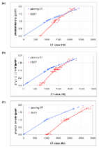
          In this study, the performance of a new iterative reconstruction algorithm, the pre-clinical AcurosXB iCBCT algorithm, has been characterized on Varian Halcyon linear accelerators with respect to the potential of radiotherapy dose calculations on CBCT images. The study utilized various phantom setups to verify the accuracy of the pre-clinical algorithm under different scatter conditions and compared dose calculations performed on CBCT images reconstructed with the pre-clinical algorithm to those performed on typical planning CT images. The results indicated that despite showing improvements compared to the existing iCBCT protocol, certain restrictions should be introduced when the pre-clinical AcurosXB iCBCT algorithm was used for dose calculations. Changes in the scatter condition exhibited a larger effect on CBCTs than on planning CTs. Therefore, users should be careful in offsetting the patient and positioning the patient’s arms if the resultant images will be used for dose calculations. In addition, protocols with different kV settings should be approached with caution, where 100 kV protocols should only be used to scan the head and neck area, while the rest of the body should be scanned with the 125 kV and 140 kV protocols. When the patient is set up properly and the appropriate energy is selected for the anatomical area, the uncertainty of using the novel AcurosXB iCBCT algorithm for treatment planning dose calculation is within ±2.0%.

          Related collections

          Most cited references22

          • Record: found
          • Abstract: found
          • Article: not found

          Quality assurance for image-guided radiation therapy utilizing CT-based technologies: a report of the AAPM TG-179.

          Commercial CT-based image-guided radiotherapy (IGRT) systems allow widespread management of geometric variations in patient setup and internal organ motion. This document provides consensus recommendations for quality assurance protocols that ensure patient safety and patient treatment fidelity for such systems.
            Bookmark
            • Record: found
            • Abstract: found
            • Article: not found

            Evaluation of on-board kV cone beam CT (CBCT)-based dose calculation.

            On-board CBCT images are used to generate patient geometric models to assist patient setup. The image data can also, potentially, be used for dose reconstruction in combination with the fluence maps from treatment plan. Here we evaluate the achievable accuracy in using a kV CBCT for dose calculation. Relative electron density as a function of HU was obtained for both planning CT (pCT) and CBCT using a Catphan-600 calibration phantom. The CBCT calibration stability was monitored weekly for 8 consecutive weeks. A clinical treatment planning system was employed for pCT- and CBCT-based dose calculations and subsequent comparisons. Phantom and patient studies were carried out. In the former study, both Catphan-600 and pelvic phantoms were employed to evaluate the dosimetric performance of the full-fan and half-fan scanning modes. To evaluate the dosimetric influence of motion artefacts commonly seen in CBCT images, the Catphan-600 phantom was scanned with and without cyclic motion using the pCT and CBCT scanners. The doses computed based on the four sets of CT images (pCT and CBCT with/without motion) were compared quantitatively. The patient studies included a lung case and three prostate cases. The lung case was employed to further assess the adverse effect of intra-scan organ motion. Unlike the phantom study, the pCT of a patient is generally acquired at the time of simulation and the anatomy may be different from that of CBCT acquired at the time of treatment delivery because of organ deformation. To tackle the problem, we introduced a set of modified CBCT images (mCBCT) for each patient, which possesses the geometric information of the CBCT but the electronic density distribution mapped from the pCT with the help of a BSpline deformable image registration software. In the patient study, the dose computed with the mCBCT was used as a surrogate of the 'ground truth'. We found that the CBCT electron density calibration curve differs moderately from that of pCT. No significant fluctuation was observed in the calibration over the period of 8 weeks. For the static phantom, the doses computed based on pCT and CBCT agreed to within 1%. A notable difference in CBCT- and pCT-based dose distributions was found for the motion phantom due to the motion artefacts which appeared in the CBCT images (the maximum discrepancy was found to be approximately 3.0% in the high dose region). The motion artefacts-induced dosimetric inaccuracy was also observed in the lung patient study. For the prostate cases, the mCBCT- and CBCT-based dose calculations yielded very close results (<2%). Coupled with the phantom data, it is concluded that the CBCT can be employed directly for dose calculation for a disease site such as the prostate, where there is little motion artefact. In the prostate case study, we also noted a large discrepancy between the original treatment plan and the CBCT (or mCBCT)-based calculation, suggesting the importance of inter-fractional organ movement and the need for adaptive therapy to compensate for the anatomical changes in the future.
              Bookmark
              • Record: found
              • Abstract: found
              • Article: found
              Is Open Access

              Investigation of the usability of conebeam CT data sets for dose calculation

              Background To investigate the feasibility and accuracy of dose calculation in cone beam CT (CBCT) data sets. Methods Kilovoltage CBCT images were acquired with the Elekta XVI system, CT studies generated with a conventional multi-slice CT scanner (Siemens Somatom Sensation Open) served as reference images. Material specific volumes of interest (VOI) were defined for commercial CT Phantoms (CATPhan® and Gammex RMI®) and CT values were evaluated in CT and CBCT images. For CBCT imaging, the influence of image acquisition parameters such as tube voltage, with or without filter (F1 or F0) and collimation on the CT values was investigated. CBCT images of 33 patients (pelvis n = 11, thorax n = 11, head n = 11) were compared with corresponding planning CT studies. Dose distributions for three different treatment plans were calculated in CT and CBCT images and differences were evaluated. Four different correction strategies to match CT values (HU) and density (D) in CBCT images were analysed: standard CT HU-D table without adjustment for CBCT; phantom based HU-D tables; patient group based HU-D tables (pelvis, thorax, head); and patient specific HU-D tables. Results CT values in the CBCT images of the CATPhan® were highly variable depending on the image acquisition parameters: a mean difference of 564 HU ± 377 HU was calculated between CT values determined from the planning CT and CBCT images. Hence, two protocols were selected for CBCT imaging in the further part of the study and HU-D tables were always specific for these protocols (pelvis and thorax with M20F1 filter, 120 kV; head S10F0 no filter, 100 kV). For dose calculation in real patient CBCT images, the largest differences between CT and CBCT were observed for the standard CT HU-D table: differences were 8.0% ± 5.7%, 10.9% ± 6.8% and 14.5% ± 10.4% respectively for pelvis, thorax and head patients using clinical treatment plans. The use of patient and group based HU-D tables resulted in small dose differences between planning CT and CBCT: 0.9% ± 0.9%, 1.8% ± 1.6%, 1.5% ± 2.5% for pelvis, thorax and head patients, respectively. The application of the phantom based HU-D table was acceptable for the head patients but larger deviations were determined for the pelvis and thorax patient populations. Conclusion The generation of three HU-D tables specific for the anatomical regions pelvis, thorax and head and specific for the corresponding CBCT image acquisition parameters resulted in accurate dose calculation in CBCT images. Once these HU-D tables are created, direct dose calculation on CBCT datasets is possible without the need of a reference CT images for pixel value calibration.
                Bookmark

                Author and article information

                Contributors
                Journal
                Biomedical Physics & Engineering Express
                Biomed. Phys. Eng. Express
                IOP Publishing
                2057-1976
                February 25 2022
                March 01 2022
                February 25 2022
                March 01 2022
                : 8
                : 2
                : 025023
                Article
                10.1088/2057-1976/ac536b
                6e7932bc-4db7-4d36-a737-77e328c1c5e7
                © 2022

                http://creativecommons.org/licenses/by/4.0/

                History

                Comments

                Comment on this article