2
views
0
recommends
+1 Recommend
0 collections
    0
    shares
      • Record: found
      • Abstract: found
      • Article: found
      Is Open Access

      Glioma: bridging the tumor microenvironment, patient immune profiles and novel personalized immunotherapy

      review-article

      Read this article at

      Bookmark
          There is no author summary for this article yet. Authors can add summaries to their articles on ScienceOpen to make them more accessible to a non-specialist audience.

          Abstract

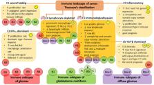
          Glioma is the most common primary brain tumor, characterized by a consistently high patient mortality rate and a dismal prognosis affecting both survival and quality of life. Substantial evidence underscores the vital role of the immune system in eradicating tumors effectively and preventing metastasis, underscoring the importance of cancer immunotherapy which could potentially address the challenges in glioma therapy. Although glioma immunotherapies have shown promise in preclinical and early-phase clinical trials, they face specific limitations and challenges that have hindered their success in further phase III trials. Resistance to therapy has been a major challenge across many experimental approaches, and as of now, no immunotherapies have been approved. In addition, there are several other limitations facing glioma immunotherapy in clinical trials, such as high intra- and inter-tumoral heterogeneity, an inherently immunosuppressive microenvironment, the unique tissue-specific interactions between the central nervous system and the peripheral immune system, the existence of the blood-brain barrier, which is a physical barrier to drug delivery, and the immunosuppressive effects of standard therapy. Therefore, in this review, we delve into several challenges that need to be addressed to achieve boosted immunotherapy against gliomas. First, we discuss the hurdles posed by the glioma microenvironment, particularly its primary cellular inhabitants, in particular tumor-associated microglia and macrophages (TAMs), and myeloid cells, which represent a significant barrier to effective immunotherapy. Here we emphasize the impact of inducing immunogenic cell death (ICD) on the migration of Th17 cells into the tumor microenvironment, converting it into an immunologically “hot” environment and enhancing the effectiveness of ongoing immunotherapy. Next, we address the challenge associated with the accurate identification and characterization of the primary immune profiles of gliomas, and their implications for patient prognosis, which can facilitate the selection of personalized treatment regimens and predict the patient’s response to immunotherapy. Finally, we explore a prospective approach to developing highly personalized vaccination strategies against gliomas, based on the search for patient-specific neoantigens. All the pertinent challenges discussed in this review will serve as a compass for future developments in immunotherapeutic strategies against gliomas, paving the way for upcoming preclinical and clinical research endeavors.

          Related collections

          Most cited references148

          • Record: found
          • Abstract: found
          • Article: found

          The 2016 World Health Organization Classification of Tumors of the Central Nervous System: a summary.

          The 2016 World Health Organization Classification of Tumors of the Central Nervous System is both a conceptual and practical advance over its 2007 predecessor. For the first time, the WHO classification of CNS tumors uses molecular parameters in addition to histology to define many tumor entities, thus formulating a concept for how CNS tumor diagnoses should be structured in the molecular era. As such, the 2016 CNS WHO presents major restructuring of the diffuse gliomas, medulloblastomas and other embryonal tumors, and incorporates new entities that are defined by both histology and molecular features, including glioblastoma, IDH-wildtype and glioblastoma, IDH-mutant; diffuse midline glioma, H3 K27M-mutant; RELA fusion-positive ependymoma; medulloblastoma, WNT-activated and medulloblastoma, SHH-activated; and embryonal tumour with multilayered rosettes, C19MC-altered. The 2016 edition has added newly recognized neoplasms, and has deleted some entities, variants and patterns that no longer have diagnostic and/or biological relevance. Other notable changes include the addition of brain invasion as a criterion for atypical meningioma and the introduction of a soft tissue-type grading system for the now combined entity of solitary fibrous tumor / hemangiopericytoma-a departure from the manner by which other CNS tumors are graded. Overall, it is hoped that the 2016 CNS WHO will facilitate clinical, experimental and epidemiological studies that will lead to improvements in the lives of patients with brain tumors.
            Bookmark
            • Record: found
            • Abstract: found
            • Article: not found

            Radiotherapy plus Concomitant and Adjuvant Temozolomide for Glioblastoma

            Glioblastoma, the most common primary brain tumor in adults, is usually rapidly fatal. The current standard of care for newly diagnosed glioblastoma is surgical resection to the extent feasible, followed by adjuvant radiotherapy. In this trial we compared radiotherapy alone with radiotherapy plus temozolomide, given concomitantly with and after radiotherapy, in terms of efficacy and safety. Patients with newly diagnosed, histologically confirmed glioblastoma were randomly assigned to receive radiotherapy alone (fractionated focal irradiation in daily fractions of 2 Gy given 5 days per week for 6 weeks, for a total of 60 Gy) or radiotherapy plus continuous daily temozolomide (75 mg per square meter of body-surface area per day, 7 days per week from the first to the last day of radiotherapy), followed by six cycles of adjuvant temozolomide (150 to 200 mg per square meter for 5 days during each 28-day cycle). The primary end point was overall survival. A total of 573 patients from 85 centers underwent randomization. The median age was 56 years, and 84 percent of patients had undergone debulking surgery. At a median follow-up of 28 months, the median survival was 14.6 months with radiotherapy plus temozolomide and 12.1 months with radiotherapy alone. The unadjusted hazard ratio for death in the radiotherapy-plus-temozolomide group was 0.63 (95 percent confidence interval, 0.52 to 0.75; P<0.001 by the log-rank test). The two-year survival rate was 26.5 percent with radiotherapy plus temozolomide and 10.4 percent with radiotherapy alone. Concomitant treatment with radiotherapy plus temozolomide resulted in grade 3 or 4 hematologic toxic effects in 7 percent of patients. The addition of temozolomide to radiotherapy for newly diagnosed glioblastoma resulted in a clinically meaningful and statistically significant survival benefit with minimal additional toxicity. Copyright 2005 Massachusetts Medical Society.
              Bookmark
              • Record: found
              • Abstract: found
              • Article: not found

              The 2021 WHO Classification of Tumors of the Central Nervous System: a summary

              The fifth edition of the WHO Classification of Tumors of the Central Nervous System (CNS), published in 2021, is the sixth version of the international standard for the classification of brain and spinal cord tumors. Building on the 2016 updated fourth edition and the work of the Consortium to Inform Molecular and Practical Approaches to CNS Tumor Taxonomy, the 2021 fifth edition introduces major changes that advance the role of molecular diagnostics in CNS tumor classification. At the same time, it remains wedded to other established approaches to tumor diagnosis such as histology and immunohistochemistry. In doing so, the fifth edition establishes some different approaches to both CNS tumor nomenclature and grading and it emphasizes the importance of integrated diagnoses and layered reports. New tumor types and subtypes are introduced, some based on novel diagnostic technologies such as DNA methylome profiling. The present review summarizes the major general changes in the 2021 fifth edition classification and the specific changes in each taxonomic category. It is hoped that this summary provides an overview to facilitate more in-depth exploration of the entire fifth edition of the WHO Classification of Tumors of the Central Nervous System.
                Bookmark

                Author and article information

                Contributors
                URI : https://loop.frontiersin.org/people/97625Role: Role: Role: Role: Role: Role: Role:
                Role: Role: Role: Role:
                URI : https://loop.frontiersin.org/people/919322Role: Role: Role: Role:
                URI : https://loop.frontiersin.org/people/655257Role: Role: Role:
                URI : https://loop.frontiersin.org/people/97620Role: Role: Role:
                URI : https://loop.frontiersin.org/people/35016Role: Role: Role:
                Journal
                Front Immunol
                Front Immunol
                Front. Immunol.
                Frontiers in Immunology
                Frontiers Media S.A.
                1664-3224
                11 January 2024
                2023
                : 14
                : 1299064
                Affiliations
                [1] 1Institute of Biology and Biomedicine, National Research Lobachevsky State University of Nizhny Novgorod , Nizhny Novgorod, Russia
                [2] 2Neuroscience Research Institute, National Research Lobachevsky State University of Nizhny Novgorod , Nizhny Novgorod, Russia
                [3] 3Cell Death Investigation and Therapy Laboratory, Anatomy and Embryology Unit, Department of Human Structure and Repair, Faculty of Medicine and Health Sciences, Ghent University , Ghent, Belgium
                [4] 4Faculty of Biology and Biotechnologies, National Research University Higher School of Economics , Moscow, Russia
                [5] 5Department of Pathophysiology, Sechenov First Moscow State Medical University (Sechenov University) , Moscow, Russia
                [6] 6Cancer Research Institute Ghent , Ghent, Belgium
                Author notes

                Edited by: Xiao Liang, Sichuan University, China

                Reviewed by: Serena Pellegatta, IRCCS Carlo Besta Neurological Institute Foundation, Italy

                Felipe J. Núñez, CONICET Institute of Biology and Experimental Medicine (IBYME), Argentina

                *Correspondence: Dmitri V. Krysko, dmitri.krysko@ 123456ugent.be

                †These authors share senior authorship

                Article
                10.3389/fimmu.2023.1299064
                10809268
                38274827
                6d2ad622-479a-40c5-b504-6fe92eed2bc5
                Copyright © 2024 Mishchenko, Turubanova, Gorshkova, Krysko, Vedunova and Krysko

                This is an open-access article distributed under the terms of the Creative Commons Attribution License (CC BY). The use, distribution or reproduction in other forums is permitted, provided the original author(s) and the copyright owner(s) are credited and that the original publication in this journal is cited, in accordance with accepted academic practice. No use, distribution or reproduction is permitted which does not comply with these terms.

                History
                : 22 September 2023
                : 11 December 2023
                Page count
                Figures: 2, Tables: 1, Equations: 0, References: 188, Pages: 16, Words: 7780
                Funding
                The author(s) declare financial support was received for the research, authorship, and/or publication of this article. The study was supported by a grant from Russian Science Foundation (RSF, project no. 22-15-00376, https://rscf.ru/en/project/22-15-00376/).
                Categories
                Immunology
                Review
                Custom metadata
                Cancer Immunity and Immunotherapy

                Immunology
                glioma,cancer immunotherapy,immunogenic cell death,icd,tumor microenvironment,th17,anti-tumor vaccination,tumor neoantigens

                Comments

                Comment on this article