0
views
0
recommends
+1 Recommend
0 collections
    0
    shares
      • Record: found
      • Abstract: found
      • Article: found
      Is Open Access

      Treatment of giant prostatic urethral stone with prostatolithotomy case report

      case-report

      Read this article at

      Bookmark
          There is no author summary for this article yet. Authors can add summaries to their articles on ScienceOpen to make them more accessible to a non-specialist audience.

          Abstract

          Introduction

          The patient with prostatic urethral stones of the size mentioned in the case report is very rare, and there is no standard surgical procedure for patients with giant stones in the prostatic urethra.

          Presentation of case

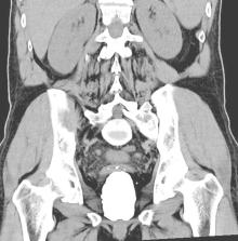
          A 62-year-old male patient presented to the emergency department with complaints of dysuria and hematuria. Computed tomography showed a prostatic urethral stone measuring 78x48x56 mm. Open prostatolithotomy was performed by extending the bladder incision towards the prostate capsule and the stone was removed.

          Discussion

          Prostate stones can be classified into two types: true prostate stones, which form within the prostate's tissues, and urethral stones, which develop in the prostatic urethra. Urethral stones can be primary (forming in the urethra) or secondary (migrating from the upper urinary tract).

          Conclusion

          Treatment options vary based on stone size and patient history, with endoscopy recommended as the primary approach. However, in cases with large stone burdens, open surgical methods may be preferred.

          Highlights

          • Giant calculi in the prostatic cavity, though uncommon, pose unique diagnostic and management challenges.

          • This case report presents a rare 78x48x56 mm prostatic urethral stone.

          • Open prostatolithotomy was successfully performed, offering an alternative to endoscopic methods for large stones.

          • This report provides valuable insights for urologists faced with similar rare and complex cases.

          Related collections

          Most cited references11

          • Record: found
          • Abstract: found
          • Article: not found

          The SCARE 2020 Guideline: Updating Consensus Surgical CAse REport (SCARE) Guidelines

          The SCARE Guidelines were first published in 2016 and were last updated in 2018. They provide a structure for reporting surgical case reports and are used and endorsed by authors, journal editors and reviewers, in order to increase robustness and transparency in reporting surgical cases. They must be kept up to date in order to drive forwards reporting quality. As such, we have updated these guidelines via a DELPHI consensus exercise.
            Bookmark
            • Record: found
            • Abstract: found
            • Article: not found

            Prostatic calculi: a review.

            Prostatic calculi are rare in children, infrequent below age 40, and common in males over 50. They may be solitary but usually occur in clusters and are associated with some other disease process (nodular hyperplasia, prostatic carcinoma, metabolic abnormalities). They are most often asymptomatic; however, symptoms that may be attributable to prostatic calculi include reduction of the urinary stream, prostatism, and intense lower back and leg pain. Treatment may be accomplished by transurethral resection, but prostatectomy is the best means to insure complete removal. Prostatic calculi may form by two related mechanisms with obstruction and stasis of prostatic fluid as central entities in both. These mechanisms are calcification of corpora amylacea and simple precipitation of prostatic secretion. They may arise spontaneously, initiating an inflammatory reaction that contributes to their growth, or they may arise as the consequence of another pathologic situation producing acinar obstruction. It appears that infection probably occurs secondary to stone formation.
              Bookmark
              • Record: found
              • Abstract: found
              • Article: not found

              Imaging of male urethra.

              The male urethral imaging and pathology is not widespread in the radiology literature because this part of the urinary tract is easily studied by urologists with clinical or endoscopic examinations. Ultrasonography and MR imaging are increasingly being used in association with voiding cystourethrography and retrograde urethrography. The posterior urethra is being studied with voiding cystourethrography or voiding sonography which allows the detection of bladder neck pathology, post-surgical stenosis, and neoplasms. The functional aspects of the bladder neck and posterior urethra can be monitored continuously in patients with neuromuscular dysfunction of the bladder. The anterior urethral anatomy and pathology is commonly explored by retrograde urethrography, but recently sonourethrography and MR imaging have been proposed, distending the lumen with simple saline solution instead of iodinated contrast media. They are being used to study the urethral mucosa and the periurethral spongy tissue which can be involved in the urethral pathologies such as strictures, diverticula, trauma, and tumors. Imaging has an important role to play in the study of the diseases of the male urethra since it can detect pathology not visible on urethroscopy. The new imaging techniques in this area, such as sonography and MR, can provide adjunct information that cannot be obtained with other modalities.
                Bookmark

                Author and article information

                Contributors
                Journal
                Int J Surg Case Rep
                Int J Surg Case Rep
                International Journal of Surgery Case Reports
                Elsevier
                2210-2612
                20 December 2023
                January 2024
                20 December 2023
                : 114
                : 109136
                Affiliations
                [a ]Manisa Merkez Efendi State Hospital, Department of Urology, Manisa, Turkey
                [b ]İzmir Foça State Hospital, Department of Urology, İzmir, Turkey
                [c ]Manisa Celal Bayar University Faculty of Medicine, Department of Urology, Manisa, Turkey
                Author notes
                [* ]Corresponding author at: Department of Medicine, Section of Urology, Manisa Merkez Efendi State Hospital, 45030 Yunusemre, Manisa, Turkey. yunusbozkurt88@ 123456hotmail.com
                Article
                S2210-2612(23)01265-8 109136
                10.1016/j.ijscr.2023.109136
                10800754
                38134615
                6ca15221-e892-422a-8155-8d31e6a58ee6
                © 2023 The Author(s)

                This is an open access article under the CC BY license (http://creativecommons.org/licenses/by/4.0/).

                History
                : 13 November 2023
                : 3 December 2023
                : 5 December 2023
                Categories
                Case Report

                urethral stone,giant stone,prostate,case report
                urethral stone, giant stone, prostate, case report

                Comments

                Comment on this article