67
views
0
recommends
+1 Recommend
0 collections
    0
    shares
      • Record: found
      • Abstract: found
      • Article: found
      Is Open Access

      Visible Perforating Lateral Osteotomy: Internal Perforating Technique with Wide Periosteal Dissection

      brief-report

      Read this article at

      Bookmark
          There is no author summary for this article yet. Authors can add summaries to their articles on ScienceOpen to make them more accessible to a non-specialist audience.

          Abstract

          There are two general categories of lateral osteotomy techniques—the external perforating method and the internal continuous method. Regardless of which technique is used, procedural effectiveness is hampered by limited visualization in the surgical field. Considering this point, we devised a new technique that involves using a wide subperiosteal dissection and internal perforation under direct visualization. Using an intranasal approach, whereby the visibility of the intended fracture line was maintained, enabled a greater degree of control, and in turn, results that were more precise, and thus predictable and reproducible. Traditionally, it has been taken as dogma that the periosteum must be preserved, considering the potential for dead space and bony instability; however, under sufficient visualization of the surgical field with an internal perforating method, complete osteotomy with fully preserved intranasal mucosa could be conducted exactly as intended. This intact mucosal lining compensates for the elevated periosteum. Compressive dressing and drainage through a Silastic angio-needle catheter enabled the elimination of dead space. Therefore, precise, reproducible, and predictable osteotomy minimizing the potential for associated complications such as ecchymosis, that is, bruising owing to hemorrhage, could be performed. In this article, we introduce a novel technique for lateral osteotomy with improved visualization.

          Related collections

          Most cited references14

          • Record: found
          • Abstract: found
          • Article: not found

          Nasal osteotomies: a clinical comparison of the perforating methods versus the continuous technique.

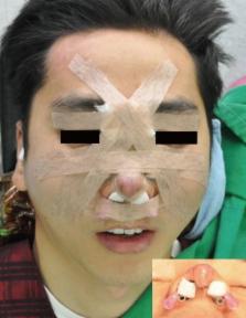
          Do perforating lateral osteotomies cause less ecchymosis and edema compared with the popular continuous method? Many studies have demonstrated that perforated osteotomies cause less trauma and periosteal disruption. Numerous investigators have subjectively perceived less postoperative ecchymosis and edema, but no clinical study has compared the perforated methods versus the continuous technique in the same patient. This prospective, randomized, partially blinded study was designed to test the hypothesis that the perforating method causes less postoperative ecchymosis and edema compared with the continuous lateral osteotomy technique. The questions remain: does the type of perforating osteotomy affect the results? Does a percutaneous approach cause more ecchymosis and edema by the access maneuver of piercing the skin? The two perforating lateral nasal osteotomy techniques require the same 2-mm straight osteotome, so any genuine difference in postoperative ecchymosis or edema could only be attributed to the differing surgical approaches. Accordingly, this study also tests whether the external percutaneous perforating osteotomy causes more ecchymosis and edema than the internal transnasal perforating method. Twenty-five consecutive rhinoplasty patients (group A) requiring bilateral osteotomies (50 total lateral osteotomies) were randomized so that each patient received an internal/transnasal perforating lateral osteotomy (2-mm straight chisel) on one side and an internal/transnasal continuous osteotomy (4-mm curved, guarded osteotome) on the other. The next 25 patients studied (group B) received an external/percutaneous perforating lateral osteotomy (same 2-mm straight chisel as used in group A) on one side and the same internal/transnasal continuous osteotomy on the other. The final 25 consecutive rhinoplasty patients (group C) received an external percutaneous perforating lateral osteotomy on one side and an internal transnasal perforating lateral osteotomy on the other. The entry sites for the perforating osteotomies were either external (groups B and C) with a percutaneous skin puncture or intranasal (groups A and C) at the pyriform aperture. All 75 patients (150 total lateral osteotomies) initialed the surgical plan on the Gunter rhinoplasty worksheet, which has been approved by the Institutional Review Board of Abbott-Northwestern Hospital, Minneapolis, Minnesota (study no. 1341-1 M). All patients were evaluated for ecchymosis and edema on the left versus the right side of the face at 2 to 3, 7, and 21 days after the operation. The clinical evaluation was performed by two blinded examiners (clinic registered nurse and the patient with his or her family) and a partially blinded examiner (the surgeon, who did not refresh his memory about the randomization). To compare the two methods in each study (groups A, B, and C) for the six outcomes (edema and ecchymosis at 2 to 3, 7, and 21 days), the authors used an exact binomial test of the null hypothesis that the treatments do not differ. To compare the two methods in each study (groups A, B, and C) using all six outcomes simultaneously, the authors used a permutation test. By both testing methods, the perforating internal method was superior to the continuous technique (group A; p < 0.01 in both tests). Although the perforating external method gave better results than the continuous technique (group B) and the perforating internal method gave better results than the perforating external method (group C), neither of these differences was significant by either testing method. A lateral osteotomy technique should be precise, reproducible, and safe, and it should minimize ecchymosis and edema. Since edema and ecchymosis are comparable regardless of osteotome size, this prospective randomized study confirms the subjective clinical impression that perforating lateral osteotomies with a 2-mm straight osteotome reduce postoperative ecchymosis and edema in rhinoplasty patients compared with the continuous osteotomy (4-mm curved, guarded osteotome). These findings should encourage te the use of perforating osteotomies rather than continuous osteotomies.
            Bookmark
            • Record: found
            • Abstract: found
            • Article: not found

            The lateral nasal osteotomy in rhinoplasty: an anatomic endoscopic comparison of the external versus the internal approach.

            A precise and reproducible lateral osteotomy is a requirement for successful rhinoplasty. Two basic techniques have evolved: the external perforated method and the internal continuous method. The literature supporting the external perforated technique maintains that it contributes to a controlled, stable fracture and produces less nasal airway narrowing, hemorrhage, edema, and ecchymosis; however, the continuous internal method is used by many rhinoplasty surgeons. Our study was designed to compare the two techniques in the fresh cadaver nose using a blinded endoscopic evaluation of the nasal mucosa after the osteotomies were performed by one of these two techniques. Nineteen fresh cadaver heads had an external perforated lateral osteotomy performed on one side and an internal continuous lateral osteotomy performed on the alternate side by an investigator with experience in the use of both osteotomies. In a blinded fashion, four different investigators used nasal endoscopy to detect mucosal perforations and bony irregularities. Eleven percent of the perforated osteotomies resulted in mucosal tearing as opposed to 74 percent of the continuous osteotomies (p < 0.001). This anatomic study confirms our clinical experience that the external perforated osteotomy results in a more controlled fracture with less intranasal trauma and can minimize the associated morbidity (hemorrhage, edema, and ecchymosis) in the rhinoplasty patient.
              Bookmark
              • Record: found
              • Abstract: not found
              • Article: not found

              An update on the lateral nasal osteotomy in rhinoplasty: an anatomic endoscopic comparison of the external versus the internal approach.

                Bookmark

                Author and article information

                Journal
                Arch Plast Surg
                Arch Plast Surg
                APS
                Archives of Plastic Surgery
                The Korean Society of Plastic and Reconstructive Surgeons
                2234-6163
                2234-6171
                January 2016
                15 January 2016
                : 43
                : 1
                : 88-92
                Affiliations
                [1 ]Glovi Plastic Surgery, Seoul, Korea.
                [2 ]Department of Plastic and Reconstructive Surgery, Soonchunhyang University Bucheon Hospital, Soonchunhyang University College of Medicine, Bucheon, Korea.
                Author notes
                Correspondence: Eun Soo Park. Department of Plastic and Reconstructive Surgery, Soonchunhyang University Bucheon Hospital, Soonchunhyang University College of Medicine, 170 Jomaru-ro, Wonmi-gu, Bucheon 14584, Korea. Tel: +82-32-621-5319, Fax: +82-32-621-5316, peunsoo@ 123456schmc.ac.kr
                Article
                10.5999/aps.2016.43.1.88
                4738135
                26848452
                69b439c0-9e83-4b5e-8c31-6bfa7b36903d
                Copyright © 2016 The Korean Society of Plastic and Reconstructive Surgeons

                This is an Open Access article distributed under the terms of the Creative Commons Attribution Non-Commercial License ( http://creativecommons.org/licenses/by-nc/3.0/), which permits unrestricted non-commercial use, distribution, and reproduction in any medium, provided the original work is properly cited.

                History
                : 02 April 2015
                : 23 September 2015
                : 19 October 2015
                Funding
                Funded by: Soonchunhyang University, CrossRef http://dx.doi.org/10.13039/501100002560;
                Categories
                Idea and Innovation

                Surgery
                rhinoplasty,osteotomy,ecchymosis,dissection
                Surgery
                rhinoplasty, osteotomy, ecchymosis, dissection

                Comments

                Comment on this article