29
views
0
recommends
+1 Recommend
0 collections
    0
    shares
      • Record: found
      • Abstract: found
      • Article: found
      Is Open Access

      Manipulations of MeCP2 in glutamatergic neurons highlight their contributions to Rett and other neurological disorders

      research-article

      Read this article at

      Bookmark
          There is no author summary for this article yet. Authors can add summaries to their articles on ScienceOpen to make them more accessible to a non-specialist audience.

          Abstract

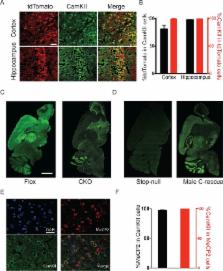
          Many postnatal onset neurological disorders such as autism spectrum disorders (ASDs) and intellectual disability are thought to arise largely from disruption of excitatory/inhibitory homeostasis. Although mouse models of Rett syndrome (RTT), a postnatal neurological disorder caused by loss-of-function mutations in MECP2, display impaired excitatory neurotransmission, the RTT phenotype can be largely reproduced in mice simply by removing MeCP2 from inhibitory GABAergic neurons. To determine what role excitatory signaling impairment might play in RTT pathogenesis, we generated conditional mouse models with Mecp2 either removed from or expressed solely in glutamatergic neurons. MeCP2 deficiency in glutamatergic neurons leads to early lethality, obesity, tremor, altered anxiety-like behaviors, and impaired acoustic startle response, which is distinct from the phenotype of mice lacking MeCP2 only in inhibitory neurons. These findings reveal a role for excitatory signaling impairment in specific neurobehavioral abnormalities shared by RTT and other postnatal neurological disorders.

          DOI: http://dx.doi.org/10.7554/eLife.14199.001

          eLife digest

          Rett syndrome is a childhood brain disorder that mainly affects girls and causes symptoms including anxiety, tremors, uncoordinated movements and breathing difficulties. Rett syndrome is caused by mutations in a gene called MECP2, which is found on the X chromosome. Males with MECP2 mutations are rare but have more severe symptoms and die young. Many researchers who study Rett syndrome use mice as a model of the disorder. In particular, male mice with the mouse equivalent of the human MECP2 gene switched off in every cell in the body (also known as Mecp2-null mice) show many of the features of Rett syndrome and die at a young age.

          The MECP2 gene is important for healthy brain activity. The brain contains two major types of neurons: excitatory neurons, which encourage other neurons to be active; and inhibitory neurons, which stop or dampen the activity of other neurons. In 2010, researchers reported that mice lacking Mecp2 in only their inhibitory neurons develop most of the same problems as those mice with no Mecp2 at all.

          Now, Meng et al. – who include two researchers involved in the 2010 study – have asked how deleting or activating Mecp2 only in excitatory neurons of mice affects Rett-syndrome-like symptoms. The experiments showed that male mice without Mecp2 in their excitatory neurons develop tremors, anxiety-like behaviors, abnormal seizure-like brain activity and severe obesity; these mice also die earlier than normal mice. Female mice lacking Mecp2 in half of their excitatory neurons (because the gene is on the X chromosome) were less affected than the males, and had normal life spans. These symptoms are different from those seen in mice missing Mecp2 only in inhibitory neurons.

          Meng et al. also found that if Mecp2 was switched on only in excitatory neurons of female mice (which are a model of human Rett syndrome patients) the mice were almost completely normal. In male mice (which show more severe symptoms), activating Mecp2 in only the excitatory neurons reduced the anxiety and tremors. These findings suggest that impaired excitatory neurons may be responsible for specific symptoms such as anxiety and tremors amongst other Rett-syndrome-like features.

          The next challenge is to explore how the loss of Mecp2 changes the activity of excitatory neurons in different brain regions. Further studies could also investigate if drugs that improve the activity of excitatory neurons can be used to treat Rett syndrome patients. Finally, in a related study, Ure et al. asked if activating Mecp2 in inhibitory neurons in otherwise Mecp2-null mice was enough to prevent some of their Rett syndrome-like symptoms.

          DOI: http://dx.doi.org/10.7554/eLife.14199.002

          Related collections

          Most cited references55

          • Record: found
          • Abstract: found
          • Article: not found

          Rett syndrome is caused by mutations in X-linked MECP2, encoding methyl-CpG-binding protein 2.

          Rett syndrome (RTT, MIM 312750) is a progressive neurodevelopmental disorder and one of the most common causes of mental retardation in females, with an incidence of 1 in 10,000-15,000 (ref. 2). Patients with classic RTT appear to develop normally until 6-18 months of age, then gradually lose speech and purposeful hand use, and develop microcephaly, seizures, autism, ataxia, intermittent hyperventilation and stereotypic hand movements. After initial regression, the condition stabilizes and patients usually survive into adulthood. As RTT occurs almost exclusively in females, it has been proposed that RTT is caused by an X-linked dominant mutation with lethality in hemizygous males. Previous exclusion mapping studies using RTT families mapped the locus to Xq28 (refs 6,9,10,11). Using a systematic gene screening approach, we have identified mutations in the gene (MECP2 ) encoding X-linked methyl-CpG-binding protein 2 (MeCP2) as the cause of some cases of RTT. MeCP2 selectively binds CpG dinucleotides in the mammalian genome and mediates transcriptional repression through interaction with histone deacetylase and the corepressor SIN3A (refs 12,13). In 5 of 21 sporadic patients, we found 3 de novo missense mutations in the region encoding the highly conserved methyl-binding domain (MBD) as well as a de novo frameshift and a de novo nonsense mutation, both of which disrupt the transcription repression domain (TRD). In two affected half-sisters of a RTT family, we found segregation of an additional missense mutation not detected in their obligate carrier mother. This suggests that the mother is a germline mosaic for this mutation. Our study reports the first disease-causing mutations in RTT and points to abnormal epigenetic regulation as the mechanism underlying the pathogenesis of RTT.
            Bookmark
            • Record: found
            • Abstract: found
            • Article: not found

            A model for neural development and treatment of Rett syndrome using human induced pluripotent stem cells.

            Autism spectrum disorders (ASD) are complex neurodevelopmental diseases in which different combinations of genetic mutations may contribute to the phenotype. Using Rett syndrome (RTT) as an ASD genetic model, we developed a culture system using induced pluripotent stem cells (iPSCs) from RTT patients' fibroblasts. RTT patients' iPSCs are able to undergo X-inactivation and generate functional neurons. Neurons derived from RTT-iPSCs had fewer synapses, reduced spine density, smaller soma size, altered calcium signaling and electrophysiological defects when compared to controls. Our data uncovered early alterations in developing human RTT neurons. Finally, we used RTT neurons to test the effects of drugs in rescuing synaptic defects. Our data provide evidence of an unexplored developmental window, before disease onset, in RTT syndrome where potential therapies could be successfully employed. Our model recapitulates early stages of a human neurodevelopmental disease and represents a promising cellular tool for drug screening, diagnosis and personalized treatment. Copyright © 2010 Elsevier Inc. All rights reserved.
              Bookmark
              • Record: found
              • Abstract: found
              • Article: not found

              A mouse Mecp2-null mutation causes neurological symptoms that mimic Rett syndrome.

              Rett syndrome (RTT) is an inherited neurodevelopmental disorder of females that occurs once in 10,000-15,000 births. Affected females develop normally for 6-18 months, but then lose voluntary movements, including speech and hand skills. Most RTT patients are heterozygous for mutations in the X-linked gene MECP2 (refs. 3-12), encoding a protein that binds to methylated sites in genomic DNA and facilitates gene silencing. Previous work with Mecp2-null embryonic stem cells indicated that MeCP2 is essential for mouse embryogenesis. Here we generate mice lacking Mecp2 using Cre-loxP technology. Both Mecp2-null mice and mice in which Mecp2 was deleted in brain showed severe neurological symptoms at approximately six weeks of age. Compensation for absence of MeCP2 in other tissues by MeCP1 (refs. 19,20) was not apparent in genetic or biochemical tests. After several months, heterozygous female mice also showed behavioral symptoms. The overlapping delay before symptom onset in humans and mice, despite their profoundly different rates of development, raises the possibility that stability of brain function, not brain development per se, is compromised by the absence of MeCP2.
                Bookmark

                Author and article information

                Contributors
                Role: Reviewing editor
                Journal
                eLife
                Elife
                eLife
                eLife
                eLife
                eLife Sciences Publications, Ltd
                2050-084X
                21 June 2016
                2016
                : 5
                : e14199
                Affiliations
                [1 ]deptDepartment of Neuroscience , Baylor College of Medicine , Houston, United States
                [2 ]deptJan and Dan Duncan Neurological Research Institute , Texas Children's Hospital , Houston, United States
                [3 ]deptDepartment of Molecular and Human Genetics , Baylor College of Medicine , Houston, United States
                [4 ]Howard Hughes Medical Institute, Baylor College of Medicine , Houston, United States
                [5 ]deptThe Cain Foundation Laboratories, Jan and Dan Duncan Neurological Research Institute , Texas Children's Hospital , Houston, United States
                [6 ]deptChildren's Nutrition Research Center, Department of Pediatrics , Baylor College of Medicine , Houston, United States
                [7 ]deptDepartment of Pediatrics , Baylor College of Medicine , Houston, United States
                [8 ]deptInterdepartmental Program in Translational Biology and Molecular Medicine , Baylor College of Medicine , Houston, United States
                [9 ]deptHuffington Center on Aging , Baylor College of Medicine , Houston, United States
                [10 ]deptDepartment of Molecular and Cellular Biology , Baylor College of Medicine , Houston, United States
                [11 ]deptBobby R Alford Department of Otolaryngology - Head and Neck Surgery , Baylor College of Medicine , Houston, United States
                [12]Howard Hughes Medical Institute, Harvard University , United States
                [13]Howard Hughes Medical Institute, Harvard University , United States
                Author notes
                [†]

                Department of Neurosciences, University of California, San Diego, United States.

                Author information
                http://orcid.org/0000-0003-3808-1769
                http://orcid.org/0000-0002-0700-3349
                Article
                14199
                10.7554/eLife.14199
                4946906
                27328325
                68556224-492c-46a2-92f1-dba0dd3a232f
                © 2016, Meng et al

                This article is distributed under the terms of the Creative Commons Attribution License, which permits unrestricted use and redistribution provided that the original author and source are credited.

                History
                : 04 January 2016
                : 01 June 2016
                Funding
                Funded by: FundRef http://dx.doi.org/10.13039/100001391, Whitehall Foundation;
                Award ID: Research Grant 2015-05-54
                Award Recipient :
                Funded by: FundRef http://dx.doi.org/10.13039/100007856, Baylor College of Medicine;
                Award ID: Curtis Hankamer Basic Research Fund
                Award Recipient :
                Funded by: FundRef http://dx.doi.org/10.13039/100000065, National Institute of Neurological Disorders and Stroke;
                Award ID: 5R01NS057819
                Award Recipient :
                Funded by: FundRef http://dx.doi.org/10.13039/100007857, Intellectual and Developmental Disabilities Research Center;
                Award ID: 1U54 HD083092
                Award Recipient :
                The funders had no role in study design, data collection and interpretation, or the decision to submit the work for publication.
                Categories
                Neuroscience
                Research Article
                Custom metadata
                2.5
                Excitatory signaling impairment contributes to neurological deficits shared by Rett syndrome and a number of postnatal neuropsychiatric disorders.

                Life sciences
                glutamatergic neurons,neurological disorders,mecp2,rett syndrome,mouse
                Life sciences
                glutamatergic neurons, neurological disorders, mecp2, rett syndrome, mouse

                Comments

                Comment on this article