10
views
0
recommends
+1 Recommend
0 collections
    0
    shares
      • Record: found
      • Abstract: found
      • Article: found
      Is Open Access

      CD40 signaling instructs chronic lymphocytic leukemia cells to attract monocytes via the CCR2 axis

      research-article

      Read this article at

      Bookmark
          There is no author summary for this article yet. Authors can add summaries to their articles on ScienceOpen to make them more accessible to a non-specialist audience.

          Abstract

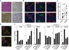
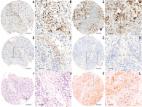
          Chronic lymphocytic leukemia (CLL) cells are provided with essential survival and proliferative signals in the lymph node microenvironment. Here, CLL cells engage in various interactions with bystander cells such as T cells and macrophages. Phenotypically distinct types of tumor infiltrating macrophages can either be tumor supportive (M2) or play a role in tumor immune surveillance (M1). Although recent in vitro findings suggest a protective role for macrophages in CLL, the actual balance between these macrophage subsets in CLL lymphoid tissue is still unclear. Furthermore, the mechanism of recruitment of monocytes towards the CLL lymph node is currently unknown. Both questions are addressed in this paper. Immunofluorescence staining of lymph node samples showed macrophage skewing towards an M2 tumor-promoting phenotype. This polarization likely results from CLL-secreted soluble factors, as both patient serum and CLL-conditioned medium recapitulated the skewing effect. Considering that CLL cell cytokine secretion is affected by adjacent T cells, we next studied CLL-mediated monocyte recruitment in the presence or absence of T-cell signals. While unstimulated CLL cells were inactive, T cell-stimulated CLL cells actively recruited monocytes. This correlated with secretion of various chemokines such as C-C-motif-ligand-2,3,4,5,7,24, C-X-C-motif-ligand-5,10, and Interleukin-10. We also identified CD40L as the responsible T-cell factor that mediated recruitment, and showed that recruitment critically depended on the C-C-motif-chemokine-receptor-2 axis. These studies show that the shaping of a tumor supportive microenvironment depends on cytokinome alterations (including C-C-motif-ligand-2) that occur after interactions between CLL, T cells and monocytes. Therefore, targeted inhibition of CD40L or C-C-motif-chemokine-receptor-2 may be relevant therapeutic options.

          Related collections

          Most cited references27

          • Record: found
          • Abstract: found
          • Article: not found

          Distinct role of macrophages in different tumor microenvironments.

          Macrophages are prominent in the stromal compartment of virtually all types of malignancy. These highly versatile cells respond to the presence of stimuli in different parts of tumors with the release of a distinct repertoire of growth factors, cytokines, chemokines, and enzymes that regulate tumor growth, angiogenesis, invasion, and/or metastasis. The distinct microenvironments where tumor-associated macrophages (TAM) act include areas of invasion where TAMs promote cancer cell motility, stromal and perivascular areas where TAMs promote metastasis, and avascular and perinecrotic areas where hypoxic TAMs stimulate angiogenesis. This review will discuss the evidence for differential regulation of TAMs in these microenvironments and provide an overview of current attempts to target or use TAMs for therapeutic purposes.
            Bookmark
            • Record: found
            • Abstract: found
            • Article: not found

            Cancer related inflammation: the macrophage connection.

            Tumor-associated macrophages (TAM) are key regulators of the link between inflammation and cancer. In the tumor microenvironment neoplastic cells shape the differentiation and functional orientation of TAM which, in turn, express several protumoral functions, including secretion of growth factors and matrix-proteases, promotion of angiogenesis and suppression of adaptive immunity. This review analyzes our current knowledge of TAM and their involvement in tumor development and progression. The interplay between TAM and neoplastic cells represents a promising target of future therapeutic approaches.
              Bookmark
              • Record: found
              • Abstract: found
              • Article: found
              Is Open Access

              Human breast cancer cells educate macrophages toward the M2 activation status

              Introduction The immune system plays a major role in cancer progression. In solid tumors, 5-40 % of the tumor mass consists of tumor-associated macrophages (TAMs) and there is usually a correlation between the number of TAMs and poor prognosis, depending on the tumor type. TAMs usually resemble M2 macrophages. Unlike M1-macrophages which have pro-inflammatory and anti-cancer functions, M2-macrophages are immunosuppressive, contribute to the matrix-remodeling, and hence favor tumor growth. The role of TAMs is not fully understood in breast cancer progression. Methods Macrophage infiltration (CD68) and activation status (HLA-DRIIα, CD163) were evaluated in a large cohort of human primary breast tumors (562 tissue microarray samples), by immunohistochemistry and scored by automated image analysis algorithms. Survival between groups was compared using the Kaplan-Meier life-table method and a Cox multivariate proportional hazards model. Macrophage education by breast cancer cells was assessed by ex vivo differentiation of peripheral blood mononuclear cells (PBMCs) in the presence or absence of breast cancer cell conditioned media (MDA-MB231, MCF-7 or T47D cell lines) and M1 or M2 inducing cytokines (respectively IFN-γ, IL-4 and IL-10). Obtained macrophages were analyzed by flow cytometry (CD14, CD16, CD64, CD86, CD200R and CD163), ELISA (IL-6, IL-8, IL-10, monocyte colony stimulating factor M-CSF) and zymography (matrix metalloproteinase 9, MMP-9). Results Clinically, we found that high numbers of CD163+ M2-macrophages were strongly associated with fast proliferation, poor differentiation, estrogen receptor negativity and histological ductal type (p<0.001) in the studied cohort of human primary breast tumors. We demonstrated ex vivo that breast cancer cell-secreted factors modulate macrophage differentiation toward the M2 phenotype. Furthermore, the more aggressive mesenchymal-like cell line MDA-MB231, which secretes high levels of M-CSF, skews macrophages toward the more immunosuppressive M2c subtype. Conclusions This study demonstrates that human breast cancer cells influence macrophage differentiation and that TAM differentiation status correlates with recurrence free survival, thus further emphasizing that TAMs can similarly affect therapy efficacy and patient outcome. Electronic supplementary material The online version of this article (doi:10.1186/s13058-015-0621-0) contains supplementary material, which is available to authorized users.
                Bookmark

                Author and article information

                Journal
                Haematologica
                Haematologica
                haematol
                Haematologica
                Haematologica
                Ferrata Storti Foundation
                0390-6078
                1592-8721
                December 2017
                29 September 2017
                : 102
                : 12
                : 2069-2076
                Affiliations
                [1 ]Department of Hematology, Academic Medical Center, University of Amsterdam; the Netherlands
                [2 ]Department of Experimental Immunology, Academic Medical Center, University of Amsterdam; the Netherlands
                [3 ]Department of Hematology, Haga Teaching Hospital, The Hague, the Netherlands
                [4 ]Lymphoma and Myeloma Center Amsterdam (LYMMCARE), the Netherlands
                Author notes
                Correspondence: a.p.kater@ 123456amc.nl

                JvB and ES contributed equally to this work as second authors, and EE and APK are senior authors

                Article
                1022069
                10.3324/haematol.2016.157206
                5709106
                28971904
                67c605d7-68fb-4aad-9b98-477d8d6201de
                Copyright© 2017 Ferrata Storti Foundation

                Material published in Haematologica is covered by copyright. All rights are reserved to the Ferrata Storti Foundation. Use of published material is allowed under the following terms and conditions:

                https://creativecommons.org/licenses/by-nc/4.0/legalcode. Copies of published material are allowed for personal or internal use. Sharing published material for non-commercial purposes is subject to the following conditions:

                https://creativecommons.org/licenses/by-nc/4.0/legalcode, sect. 3. Reproducing and sharing published material for commercial purposes is not allowed without permission in writing from the publisher.

                History
                : 27 September 2016
                : 22 September 2017
                Categories
                Article
                Chronic Lymphocytic Leukemia

                Comments

                Comment on this article