7
views
0
recommends
+1 Recommend
0 collections
    0
    shares
      • Record: found
      • Abstract: found
      • Article: found
      Is Open Access

      Histopathologic analysis of gingival lesions: A 20-year retrospective study at one academic dental center

      research-article
      1 , 2 ,
      Journal of Clinical and Experimental Dentistry
      Medicina Oral S.L.

      Read this article at

      Bookmark
          There is no author summary for this article yet. Authors can add summaries to their articles on ScienceOpen to make them more accessible to a non-specialist audience.

          Abstract

          Background

          The gingiva is part of the periodontium supporting structures surrounding the teeth and commonly involved in gingival and periodontal conditions. Assessing the distribution of gingival lesions is important for evaluating the prevalence of periodontal disease in the population to optimize the oral health care services. The purpose of this study is to report the frequency and distribution of gingival lesions biopsied from 1996–2016.

          Material and Methods

          This cross-sectional retrospective study retrieved data from all gingival lesions biopsied from 1996–2016 and sent to the King Abdulaziz University Dental Hospital oral pathology laboratory. Histologic sections were reviewed in a blinded manner by a certified oral pathologist to confirm the initial histologic diagnosis.

          Results

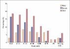
          Of the 1,248 oral-maxillofacial lesions, 119 (9.5%) gingival lesions were diagnosed. The mean age was 41.58 years. Gingival lesions were more prevalent in female patients than male patients (53.8%). The most common diagnoses were reactive lesions (41.2%). Pyogenic granuloma was the predominant lesion in the category (n=26, 21.8%), and followed by inflammatory conditions (24.4%), benign neoplasm (9.2%), malignant neoplasm (7.6%), epithelial lesions (7.6%), miscellaneous (5%), and immune-mediated diseases (5%). Squamous cell carcinoma was the only malignant neoplasm reported (7.6%; mean age, 57.44 years) and more common in male than female patients (2:1). Most biopsies were sent from oral and maxillofacial surgeons (55.6%) followed by general dentists (22.2%) and periodontists (12.8%).

          Conclusions

          Pyogenic granuloma was the most common gingival lesion. Squamous cell carcinoma was the only malignant lesion in which histologic examination was the definitive diagnostic measure. This study provides information about the frequencies and distributions of gingival lesions over 20 years.

          Key words:Gingival biopsies, retrospective, reactive lesions, oral pathology.

          Related collections

          Most cited references15

          • Record: found
          • Abstract: found
          • Article: not found

          Reactive lesions of the gingiva. A clinicopathological study of 741 cases.

          A series of 741 consecutive cases of localized hyperplastic lesions of the gingiva were studied. The lesions were reclassified into four groups: pyogenic granuloma, peripheral giant cell granuloma, fibrous hyperplasia and peripheral fibroma with calcification. This study indicates that there are some differences between these groups in age and sex distribution as well as in location and size of the lesion. Fibrous hyperplasia was the most common type, followed in descending order by pyogenic granuloma, peripheral fibroma with calcification and peripheral giant cell granuloma. The peripheral giant cell granuloma showed no sex predilection while fibrous hyperplasia, pyogenic granuloma and peripheral fibroma with calcification were more common in females. Pyogenic granuloma and peripheral fibroma with calcification occur in younger patients more often than fibrous hyperplasia, and thus may represent a stage in the development of fibrous hyperplasia.
            Bookmark
            • Record: found
            • Abstract: found
            • Article: not found

            Gingival enlargements: Differential diagnosis and review of literature.

            Gingival enlargement is one of the frequent features of gingival diseases. However due to their varied presentations, the diagnosis of these entities becomes challenging for the clinician. They can be categorized based on their etiopathogenesis, location, size, extent, etc. Based on the existing knowledge and clinical experience, a differential diagnosis can be formulated. Subsequently, after detailed investigation, clinician makes a final diagnosis or diagnosis of exclusion. A perfect diagnosis is critically important, since the management of these lesions and prevention of their recurrence is completely dependent on it. Furthermore, in some cases where gingival enlargement could be the primary sign of potentially lethal systemic diseases, a correct diagnosis of these enlargements could prove life saving for the patient or at least initiate early treatment and improve the quality of life. The purpose of this review article is to highlight significant findings of different types of gingival enlargement which would help clinician to differentiate between them. A detailed decision tree is also designed for the practitioners, which will help them arrive at a diagnosis in a systematic manner. There still could be some lesions which may present in an unusual manner and make the diagnosis challenging. By knowing the existence of common and rare presentations of gingival enlargement, one can keep a broad view when formulating a differential diagnosis of localized (isolated, discrete, regional) or generalized gingival enlargement.
              Bookmark
              • Record: found
              • Abstract: found
              • Article: found
              Is Open Access

              Prevalence of oral lesions among Saudi dental patients

              BACKGROUND AND OBJECTIVES: Few studies have been conducted in the Saudi population on oral mucosal lesions. The purpose of this study was to evaluate the type and extent of oral lesions in a study among dental patients at a college of dentistry in Saudi Arabia. PATIENTS AND METHODS: Over a 3-year period, 2552 dental outpatients were interviewed and investigated clinically for the presence of oral mucosal conditions. A thorough oral clinical examination was performed, including a radiographic examination. The diagnosis was confirmed histopathologically when necessary. RESULTS: Of 383 (15.0%) patients found to have oral mucosal lesions, females constituted 57.7% (n=221) and males 42.3% (n=162). The age range of the patients was between 15 to 73 years with a mean age of 38.2 years. The most commonly affected age group was 31 to 40 years, which comprised 21.4% (n=82) of all affected individuals. The least affected age group were individuals older than 61 years. The most common lesion was Fordyce granules (3.8%; n=98), followed by leukoedema (3.4%; n=86) and traumatic lesions (ulcer, erosion) in 1.9% (n=48). Tongue abnormalities were present in 4.0% (n=101) of all oral conditions observed, ranging from 1.4% (n=36) for fissured tongue to 0.1% (n=2) for bifid tongue. Other findings detected were torous platinus (1.3%; n=34), mandibular tori (0.1%; n=2) aphthous ulcer (0.4%; n=10), herpes simplex (0.3%; n=7), frictional hyperkeratosis (0.9%; n=23), melanosis (0.6%; n=14), lichen planus (0.3%; n=9) and nicotinic stomatitis (0.5%; n=13). CONCLUSION: The findings of this study provide information on the types and prevalence of oral lesions among Saudi dental patients. This provides baseline data for future studies about the prevalence of oral lesions in the general population.
                Bookmark

                Author and article information

                Journal
                J Clin Exp Dent
                J Clin Exp Dent
                Medicina Oral S.L.
                Journal of Clinical and Experimental Dentistry
                Medicina Oral S.L.
                1989-5488
                1 June 2018
                June 2018
                : 10
                : 6
                : e561-e566
                Affiliations
                [1 ]BDS, DSc, DABP, Assistant Professor, Periodontology Department, King Abdulaziz University, Faculty of Dentistry, Jeddah, Saudi Arabia
                [2 ]BDS, MBA, PhD, Assistant Professor, Oral Diagnostic Sciences Department, King Abdulaziz University, Faculty of Dentistry, Jeddah, Saudi Arabia
                Author notes
                Oral Diagnostic Sciences Department King Abdulaziz University Faculty of Dentistry, Jeddah, Saudi Arabia , E-mail: nmadi@ 123456kau.edu.sa

                Conflict of interest statement: This manuscript has not been published or presented elsewhere in part or in entirety and is not under consideration by another journal. We have read and understood your journal’s policies, and we believe that neither the manuscript nor the study violates any of these. The authors have no conflicts of interest.

                Article
                54766
                10.4317/jced.54766
                6005087
                29930774
                67473188-4ebc-4f2d-8344-8771dfa84279
                Copyright: © 2018 Medicina Oral S.L.

                This is an open-access article distributed under the terms of the Creative Commons Attribution License, which permits unrestricted use, distribution, and reproduction in any medium, provided the original work is properly cited.

                History
                : 26 April 2018
                : 25 February 2018
                Categories
                Research
                Oral Medicine and Pathology

                Comments

                Comment on this article