68
views
0
recommends
+1 Recommend
0 collections
    0
    shares
      • Record: found
      • Abstract: found
      • Article: found
      Is Open Access

      Pericyte-specific expression of PDGF beta receptor in mouse models with normal and deficient PDGF beta receptor signaling

      research-article
      1 , 1 , 1 ,
      Molecular Neurodegeneration
      BioMed Central

      Read this article at

      Bookmark
          There is no author summary for this article yet. Authors can add summaries to their articles on ScienceOpen to make them more accessible to a non-specialist audience.

          Abstract

          Background

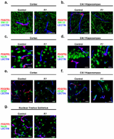
          Pericytes are integral members of the neurovascular unit. Using mouse models lacking endothelial-secreted platelet derived growth factor-B (PDGF-B) or platelet derived growth factor receptor beta (PDGFRβ) on pericytes, it has been demonstrated that PDGF-B/PDGFRβ interactions mediate pericyte recruitment to the vessel wall in the embryonic brain regulating the development of the cerebral microcirculation and the blood-brain barrier (BBB). Relatively little is known, however, about the roles of PDGF-B/PDGFRβ interactions and pericytes in the adult brain in part due to a lack of adequate and/or properly characterized experimental models. To address whether genetic disruption of PDGFRβ signaling would result in a pericyte-specific insult in adult mice, we studied the pattern and cellular distribution of PDGFRβ expression in the brain in adult control mice and F7 mice that express two hypomorphic Pdgfrβ alleles containing seven point mutations in the cytoplasmic domain of PDGFRβ that impair downstream PDGFRβ receptor signaling.

          Results

          Using dual fluorescent in situ hybridization, immunofluorescent staining for different cell types in the neurovascular unit, and a fluorescent in situ proximity ligation assay to visualize molecular PDGF-B/PDGFRβ interactions on brain tissue sections, we show for the first time that PDGFRβ is exclusively expressed in pericytes, and not in neurons, astrocytes or endothelial cells, in the adult brain of control 129S1/SvlmJ mice. PDGFRβ co-localized only with well-established pericyte markers such as Chondroitin Sulfate Proteoglycan NG2 and the xLacZ4 transgenic reporter. We next confirm pericyte-specific PDGFRβ expression in the brains of F7 mutants and show that these mice are viable in spite of substantial 40-60% reductions in regional pericyte coverage of brain capillaries.

          Conclusions

          Our data show that PDGFRβ is exclusively expressed in pericytes in the adult 129S1/Sv1mJ and F7 mouse brain. Moreover, our findings suggest that genetic disruption of PDGFRβ signaling results in a pericyte-specific insult in adult F7 mutants and will not exert a primary effect on neurons because PDGFRβ is not expressed in neurons of the adult 129S1/SvlmJ and F7 mouse brain. Therefore, mouse models with normal and deficient PDGFRβ signaling on a 129S1/SvlmJ background may effectively be used to deduce the specific roles of pericytes in maintaining the cerebral microcirculation and BBB integrity in the adult and aging brain as well as during neurodegenerative and brain vascular disorders.

          Related collections

          Most cited references30

          • Record: found
          • Abstract: found
          • Article: not found

          Pericyte loss and microaneurysm formation in PDGF-B-deficient mice.

          Platelet-derived growth factor (PDGF)-B-deficient mouse embryos were found to lack microvascular pericytes, which normally form part of the capillary wall, and they developed numerous capillary microaneurysms that ruptured at late gestation. Endothelial cells of the sprouting capillaries in the mutant mice appeared to be unable to attract PDGF-Rbeta-positive pericyte progenitor cells. Pericytes may contribute to the mechanical stability of the capillary wall. Comparisons made between PDGF null mouse phenotypes suggest a general role for PDGFs in the development of myofibroblasts.
            Bookmark
            • Record: found
            • Abstract: found
            • Article: not found

            Endothelial-mural cell signaling in vascular development and angiogenesis.

            Mural cells are essential components of blood vessels and are necessary for normal development, homeostasis, and organ function. Alterations in mural cell density or the stable attachment of mural cells to the endothelium is associated with several human diseases such as diabetic retinopathy, venous malformation, and hereditary stroke. In addition mural cells are implicated in regulating tumor growth and have thus been suggested as potential antiangiogenic targets in tumor therapy. In recent years our knowledge of mural cell function and endothelial-mural cell signaling has increased dramatically, and we now begin to understand the mechanistic basis of the key signaling pathways involved. This is mainly thanks to sophisticated in vivo experiments using a broad repertoire of genetic technologies. In this review, we summarize the five currently best understood signaling pathways implicated in mural cell biology. We discuss PDGFB/PDGFRbeta- dependent pericyte recruitment, as well as the role of angiopoietins and Tie receptors in vascular maturation. In addition, we highlight the effects of sphingosine-1-phosphate signaling on adherens junction assembly and vascular stability, as well as the role of TGF-beta-signaling in mural cell differentiation. We further reflect recent data suggesting an important function for Notch3 signaling in mural cell maturation.
              Bookmark
              • Record: found
              • Abstract: found
              • Article: not found

              Characterizing proteins and their interactions in cells and tissues using the in situ proximity ligation assay.

              The activity of proteins is typically regulated by secondary modifications and by interactions with other partners, resulting in the formation of protein complexes whose functions depend on the participating proteins. Accordingly, it is of central importance to monitor the presence of interaction complexes as well as their localization, thus providing information about the types of cells where the proteins are located and in what sub-cellular compartment these interactions occur. Several methods for visualizing protein interactions in situ have been developed during the last decade. These methods in most cases involve genetic constructs, and they have been successfully used in assays of living cell maintained in tissue culture, but they cannot easily be implemented in studies of clinical specimens. For such samples, affinity reagents like antibodies can be used to target the interacting proteins. In this review we will describe the in situ proximity ligation assays (in situ PLA), a method that is suitable for visualizing protein interactions in both tissue sections and in vitro cell lines, and we discuss research tasks when this or other method may be selected.
                Bookmark

                Author and article information

                Journal
                Mol Neurodegener
                Molecular Neurodegeneration
                BioMed Central
                1750-1326
                2010
                25 August 2010
                : 5
                : 32
                Affiliations
                [1 ]Center for Neurodegenerative and Vascular Brain Disorders, Department of Neurosurgery, University of Rochester, Rochester, NY, USA
                Article
                1750-1326-5-32
                10.1186/1750-1326-5-32
                2936891
                20738866
                61fbf4e3-823d-4e15-8afb-371090e6a658
                Copyright ©2010 Winkler et al; licensee BioMed Central Ltd.

                This is an Open Access article distributed under the terms of the Creative Commons Attribution License ( http://creativecommons.org/licenses/by/2.0), which permits unrestricted use, distribution, and reproduction in any medium, provided the original work is properly cited.

                History
                : 22 July 2010
                : 25 August 2010
                Categories
                Research Article

                Neurosciences
                Neurosciences

                Comments

                Comment on this article