52
views
0
recommends
+1 Recommend
0 collections
    0
    shares
      • Record: found
      • Abstract: found
      • Article: found
      Is Open Access

      Cone beam computed tomography: basics and applications in dentistry

      review-article

      Read this article at

      Bookmark
          There is no author summary for this article yet. Authors can add summaries to their articles on ScienceOpen to make them more accessible to a non-specialist audience.

          Abstract

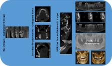
          The introduction of cone beam computed tomography (CBCT) devices, changed the way oral and maxillofacial radiology is practiced. CBCT was embraced into the dental settings very rapidly due to its compact size, low cost, low ionizing radiation exposure when compared to medical computed tomography. Alike medical CT, 3 dimensional evaluation of the maxillofacial region with minimal distortion is offered by the CBCT. This article provides an overview of basics of CBCT technology and reviews the specific application of CBCT technology to oral and maxillofacial region with few illustrations.

          Related collections

          Most cited references58

          • Record: found
          • Abstract: found
          • Article: not found

          Clinical applications of cone-beam computed tomography in dental practice.

          Cone-beam computed tomography (CBCT) systems have been designed for imaging hard tissues of the maxillofacial region. CBCT is capable of providing sub-millimetre resolution in images of high diagnostic quality, with short scanning times (10-70 seconds) and radiation dosages reportedly up to 15 times lower than those of conventional CT scans. Increasing availability of this technology provides the dental clinician with an imaging modality capable of providing a 3-dimensional representation of the maxillofacial skeleton with minimal distortion. This article provides an overview of currently available maxillofacial CBCT systems and reviews the specific application of various CBCT display modes to clinical dental practice.
            Bookmark
            • Record: found
            • Abstract: found
            • Article: not found

            What is cone-beam CT and how does it work?

            This article on x-ray cone-beam CT (CBCT) acquisition provides an overview of the fundamental principles of operation of this technology and the influence of geometric and software parameters on image quality and patient radiation dose. Advantages of the CBCT system and a summary of the uses and limitations of the images produced are discussed. All current generations of CBCT systems provide useful diagnostic images. Future enhancements most likely will be directed toward reducing scan time; providing multimodal imaging; improving image fidelity, including soft tissue contrast; and incorporating task-specific protocols to minimize patient dose.
              Bookmark
              • Record: found
              • Abstract: not found
              • Article: not found

              A new volumetric CT machine for dental imaging based on the cone-beam technique: preliminary results

                Bookmark

                Author and article information

                Journal
                J Istanb Univ Fac Dent
                J Istanb Univ Fac Dent
                J. Istanb. Univ. Fac. Dent.
                jiufd
                jiufd
                IUFD
                Journal of Istanbul University Faculty of Dentistry
                Istanbul University Faculty of Dentisty (Istanbul, Turkey )
                2149-2352
                2149-4592
                2017
                02 December 2017
                : 51
                : 3 Suppl 1
                : S102-S121
                Affiliations
                [1 ]Department of Oral, Basic and Clinical Sciences, College of Dentistry, Qassim Private Colleges, Kingdom of Saudi Arabia
                [2 ]Sandor Lifesciences Pvt. Ltd. Hyderabad, India
                Author notes
                [* ]To whom correspondence should be addressed: Dr. Elluru Venkatesh eduDept. of Oral, Basic and Clinical Sciences, College of Dentistry, Qassim Private Colleges, Kingdom of Saudi Arabia Phone:00966531696618 dr_eva1@ 123456yahoo.com
                Article
                jiufd-51-3-s498
                10.17096/jiufd.00289
                5750833
                29354314
                608b0160-9054-411c-945d-b85ded93943b
                Copyright © 2017 Journal of Istanbul University Faculty of Dentistry

                This article is licensed under Creative Commons License Attribution-NonCommercial-NoDerivatives 4.0 International (CC BY-NC-ND 4.0) license ( https://creativecommons.org/licenses/by-nc-nd/4.0/). Users must give appropriate credit, provide a link to the license, and indicate if changes were made. Users may do so in any reasonable manner, but not in any way that suggests the journal endorses its use. The material cannot be used for commercial purposes. If the user remixes, transforms, or builds upon the material, he/she may not distribute the modified material. No warranties are given. The license may not give the user all of the permissions necessary for his/her intended use. For example, other rights such as publicity, privacy, or moral rights may limit how the material can be used.

                History
                : 31 July 2017
                : 11 September 2017
                Categories
                Articles
                Biological Sciences
                Dentistry

                cone beam computed tomography,dentistry,radiography,x-ray,oral and maxillofacial imaging

                Comments

                Comment on this article