28
views
0
recommends
+1 Recommend
0 collections
    0
    shares
      • Record: found
      • Abstract: found
      • Article: found
      Is Open Access

      Pom1 regulates the assembly of Cdr2–Mid1 cortical nodes for robust spatial control of cytokinesis

      research-article

      Read this article at

      Bookmark
          There is no author summary for this article yet. Authors can add summaries to their articles on ScienceOpen to make them more accessible to a non-specialist audience.

          Abstract

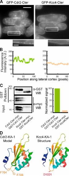
          Pom1 regulation of Cdr2 membrane association and interaction with Mid1 prevents Cdr2 assembly into stable nodes in the cell tip region, which ensures proper positioning of cytokinetic ring precursors and accurate division plane positioning in fission yeast.

          Abstract

          Proper division plane positioning is essential to achieve faithful DNA segregation and to control daughter cell size, positioning, or fate within tissues. In Schizosaccharomyces pombe, division plane positioning is controlled positively by export of the division plane positioning factor Mid1/anillin from the nucleus and negatively by the Pom1/DYRK (dual-specificity tyrosine-regulated kinase) gradients emanating from cell tips. Pom1 restricts to the cell middle cortical cytokinetic ring precursor nodes organized by the SAD-like kinase Cdr2 and Mid1/anillin through an unknown mechanism. In this study, we show that Pom1 modulates Cdr2 association with membranes by phosphorylation of a basic region cooperating with the lipid-binding KA-1 domain. Pom1 also inhibits Cdr2 interaction with Mid1, reducing its clustering ability, possibly by down-regulation of Cdr2 kinase activity. We propose that the dual regulation exerted by Pom1 on Cdr2 prevents Cdr2 assembly into stable nodes in the cell tip region where Pom1 concentration is high, which ensures proper positioning of cytokinetic ring precursors at the cell geometrical center and robust and accurate division plane positioning.

          Related collections

          Most cited references59

          • Record: found
          • Abstract: found
          • Article: not found

          Membrane recognition by phospholipid-binding domains.

          Many different globular domains bind to the surfaces of cellular membranes, or to specific phospholipid components in these membranes, and this binding is often tightly regulated. Examples include pleckstrin homology and C2 domains, which are among the largest domain families in the human proteome. Crystal structures, binding studies and analyses of subcellular localization have provided much insight into how members of this diverse group of domains bind to membranes, what features they recognize and how binding is controlled. A full appreciation of these processes is crucial for understanding how protein localization and membrane topography and trafficking are regulated in cells.
            Bookmark
            • Record: found
            • Abstract: found
            • Article: not found

            Cytokinesis in animal cells.

            Cytokinesis, the final step in cell division, partitions the contents of a single cell into two. In animal cells, cytokinesis occurs through cortical remodeling orchestrated by the anaphase spindle. Cytokinesis relies on a tight interplay between signaling and cellular mechanics and has attracted the attention of both biologists and physicists for more than a century. In this review, we provide an overview of four topics in animal cell cytokinesis: (a) signaling between the anaphase spindle and cortex, (b) the mechanics of cortical remodeling, (c) abscission, and (d) regulation of cytokinesis by the cell cycle machinery. We report on recent progress in these areas and highlight some of the outstanding questions that these findings bring into focus.
              Bookmark
              • Record: found
              • Abstract: found
              • Article: not found

              Negative regulation of mitosis by wee1+, a gene encoding a protein kinase homolog.

              Fission yeast wee1- mutants initiate mitosis at half the cell size of wild type. The wee1+ activity is required to prevent lethal premature mitosis in cells that overproduce the mitotic inducer cdc25+. This lethal phenotype was used to clone wee1+ by complementation. When wee1+ expression is increased, mitosis is delayed until cells grow to a larger size. Thus wee1+ functions as a dose-dependent inhibitor of mitosis, the first such element to be specifically identified and cloned. The carboxy-terminal region of the predicted 112 kd wee1+ protein contains protein kinase consensus sequences, suggesting that negative regulation of mitosis involves protein phosphorylation. Genetic evidence indicates that wee1+ and cdc25+ compete in a control system regulating the cdc2+ protein kinase, which is required for mitotic initiation.
                Bookmark

                Author and article information

                Journal
                J Cell Biol
                J. Cell Biol
                jcb
                jcb
                The Journal of Cell Biology
                The Rockefeller University Press
                0021-9525
                1540-8140
                7 July 2014
                : 206
                : 1
                : 61-77
                Affiliations
                [1 ]Centre de Recherche and [2 ]Laboratory of Mass Spectrometry and Proteomics, Institut Curie, F-75248 Paris, France
                [3 ]Centre National de la Recherche Scientifique, Unite Mixte de Recherche 144, F-75248 Paris, France
                [4 ]Department of Fundamental Microbiology, University of Lausanne, Lausanne CH-1015, Switzerland
                [5 ]Wellcome Trust Centre for Cell Biology, University of Edinburgh, Edinburgh EH9 3JR, Scotland, UK
                Author notes
                Correspondence to Anne Paoletti: anne.paoletti@ 123456curie.fr

                C. Bicho’s present address is Dept. of Cell and Tissue Biology, University of California, San Francisco Medical Center, San Francisco, CA 94143.

                Article
                201311097
                10.1083/jcb.201311097
                4085711
                24982431
                6061cb4a-7722-40e7-ac9a-5d886cbf5521
                © 2014 Rincon et al.

                This article is distributed under the terms of an Attribution–Noncommercial–Share Alike–No Mirror Sites license for the first six months after the publication date (see http://www.rupress.org/terms). After six months it is available under a Creative Commons License (Attribution–Noncommercial–Share Alike 3.0 Unported license, as described at http://creativecommons.org/licenses/by-nc-sa/3.0/).

                History
                : 25 November 2013
                : 28 May 2014
                Categories
                Research Articles
                Article

                Cell biology
                Cell biology

                Comments

                Comment on this article