55
views
0
recommends
+1 Recommend
0 collections
    0
    shares
      • Record: found
      • Abstract: found
      • Article: found
      Is Open Access

      Inflammation and diabetic retinal microvascular complications

      review-article

      Read this article at

      Bookmark
          There is no author summary for this article yet. Authors can add summaries to their articles on ScienceOpen to make them more accessible to a non-specialist audience.

          Abstract

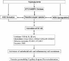
          Diabetic retinopathy (DR) is one of the most common complications of diabetes and is a leading cause of blindness in people of the working age in Western countries. A major pathology of DR is microvascular complications such as non-perfused vessels, microaneurysms, dot/blot hemorrhages, cotton-wool spots, venous beading, vascular loops, vascular leakage and neovascularization. Multiple mechanisms are involved in these alternations. This review will focus on the role of inflammation in diabetic retinal microvascular complications and discuss the potential therapies by targeting inflammation.

          Related collections

          Most cited references82

          • Record: found
          • Abstract: found
          • Article: not found

          A central role for inflammation in the pathogenesis of diabetic retinopathy.

          Diabetic retinopathy is a leading cause of adult vision loss and blindness. Much of the retinal damage that characterizes the disease results from retinal vascular leakage and nonperfusion. Diabetic retinal vascular leakage, capillary nonperfusion, and endothelial cell damage are temporary and spatially associated with retinal leukocyte stasis in early experimental diabetes. Retinal leukostasis increases within days of developing diabetes and correlates with the increased expression of retinal intercellular adhesion molecule-1 (ICAM-1) and CD18. Mice deficient in the genes encoding for the leukocyte adhesion molecules CD18 and ICAM-1 were studied in two models of diabetic retinopathy with respect to the long-term development of retinal vascular lesions. CD18-/- and ICAM-1-/- mice demonstrate significantly fewer adherent leukocytes in the retinal vasculature at 11 and 15 months after induction of diabetes with STZ. This condition is associated with fewer damaged endothelial cells and lesser vascular leakage. Galactosemia of up to 24 months causes pericyte and endothelial cell loss and formation of acellular capillaries. These changes are significantly reduced in CD18- and ICAM-1-deficient mice. Basement membrane thickening of the retinal vessels is increased in long-term galactosemic animals independent of the genetic strain. Here we show that chronic, low-grade subclinical inflammation is responsible for many of the signature vascular lesions of diabetic retinopathy. These data highlight the central and causal role of adherent leukocytes in the pathogenesis of diabetic retinopathy. They also underscore the potential utility of anti-inflammatory treatment in diabetic retinopathy.
            Bookmark
            • Record: found
            • Abstract: found
            • Article: not found

            Molecular mechanisms of angiotensin II-mediated mitochondrial dysfunction: linking mitochondrial oxidative damage and vascular endothelial dysfunction.

            Mitochondrial dysfunction is a prominent feature of most cardiovascular diseases. Angiotensin (Ang) II is an important stimulus for atherogenesis and hypertension; however, its effects on mitochondrial function remain unknown. We hypothesized that Ang II could induce mitochondrial oxidative damage that in turn might decrease endothelial nitric oxide (NO.) bioavailability and promote vascular oxidative stress. The effect of Ang II on mitochondrial ROS, mitochondrial respiration, membrane potential, glutathione, and endothelial NO. was studied in isolated mitochondria and intact bovine aortic endothelial cells using electron spin resonance, dihydroethidium high-performance liquid chromatography -based assay, Amplex Red and cationic dye fluorescence. Ang II significantly increased mitochondrial H2O2 production. This increase was blocked by preincubation of intact cells with apocynin (NADPH oxidase inhibitor), uric acid (scavenger of peroxynitrite), chelerythrine (protein kinase C inhibitor), N(G)-nitro-L-arginine methyl ester (nitric oxide synthase inhibitor), 5-hydroxydecanoate (mitochondrial ATP-sensitive potassium channels inhibitor), or glibenclamide. Depletion of p22(phox) subunit of NADPH oxidase with small interfering RNA also inhibited Ang II-mediated mitochondrial ROS production. Ang II depleted mitochondrial glutathione, increased state 4 and decreased state 3 respirations, and diminished mitochondrial respiratory control ratio. These responses were attenuated by apocynin, 5-hydroxydecanoate, and glibenclamide. In addition, 5-hydroxydecanoate prevented the Ang II-induced decrease in endothelial NO. and mitochondrial membrane potential. Therefore, Ang II induces mitochondrial dysfunction via a protein kinase C-dependent pathway by activating the endothelial cell NADPH oxidase and formation of peroxynitrite. Furthermore, mitochondrial dysfunction in response to Ang II modulates endothelial NO. and generation, which in turn has ramifications for development of endothelial dysfunction.
              Bookmark
              • Record: found
              • Abstract: found
              • Article: not found

              Increased dietary intake of omega-3-polyunsaturated fatty acids reduces pathological retinal angiogenesis.

              Many sight-threatening diseases have two critical phases, vessel loss followed by hypoxia-driven destructive neovascularization. These diseases include retinopathy of prematurity and diabetic retinopathy, leading causes of blindness in childhood and middle age affecting over 4 million people in the United States. We studied the influence of omega-3- and omega-6-polyunsaturated fatty acids (PUFAs) on vascular loss, vascular regrowth after injury, and hypoxia-induced pathological neovascularization in a mouse model of oxygen-induced retinopathy. We show that increasing omega-3-PUFA tissue levels by dietary or genetic means decreased the avascular area of the retina by increasing vessel regrowth after injury, thereby reducing the hypoxic stimulus for neovascularization. The bioactive omega-3-PUFA-derived mediators neuroprotectinD1, resolvinD1 and resolvinE1 also potently protected against neovascularization. The protective effect of omega-3-PUFAs and their bioactive metabolites was mediated, in part, through suppression of tumor necrosis factor-alpha. This inflammatory cytokine was found in a subset of microglia that was closely associated with retinal vessels. These findings indicate that increasing the sources of omega-3-PUFA or their bioactive products reduces pathological angiogenesis. Western diets are often deficient in omega-3-PUFA, and premature infants lack the important transfer from the mother to the infant of omega-3-PUFA that normally occurs in the third trimester of pregnancy. Supplementing omega-3-PUFA intake may be of benefit in preventing retinopathy.
                Bookmark

                Author and article information

                Journal
                J Cardiovasc Dis Res
                JCDR
                Journal of Cardiovascular Disease Research
                Medknow Publications Pvt Ltd (India )
                0975-3583
                0976-2833
                Apr-Jun 2011
                : 2
                : 2
                : 96-103
                Affiliations
                [1 ] Vascular Biology Center, Georgia Health Sciences University, Augusta, Georgia, USA
                [2 ] Vision Discovery Institute, Georgia Health Sciences University, Augusta, Georgia, USA
                [3 ] Department of Pharmacology and Toxicology, Georgia Health Sciences University, Augusta, Georgia, USA
                [4 ] Department of Oral Biology and Anatomy, Georgia Health Sciences University, Augusta; VA Medical Center, Augusta, Georgia, USA
                [5 ] Department of Cellular Biology and Anatomy, Georgia Health Sciences University, Augusta; VA Medical Center, Augusta, Georgia, USA
                [6 ] Department of Ophthalmology, Georgia Health Sciences University, Augusta; VA Medical Center, Augusta, Georgia, USA
                [7 ] VA Medical Center, Augusta, Georgia, USA
                Author notes
                Address for correspondence: Dr. Wenbo Zhang, Vascular Biology Center, Georgia Health Sciences University, Augusta, Georgia 30912-2500, USA. E-mail: wezhang@ 123456georgiahealth.edu
                Article
                JCDR-2-96
                10.4103/0975-3583.83035
                3144626
                21814413
                5fc8e1d2-5d72-4b0a-a89a-f6ecb8d0770f
                © Journal of Cardiovascular Disease Research

                This is an open-access article distributed under the terms of the Creative Commons Attribution-Noncommercial-Share Alike 3.0 Unported, which permits unrestricted use, distribution, and reproduction in any medium, provided the original work is properly cited.

                History
                Categories
                Invited Review

                Cardiovascular Medicine
                diabetic retinopathy,microvascular complications,inflammation
                Cardiovascular Medicine
                diabetic retinopathy, microvascular complications, inflammation

                Comments

                Comment on this article