46
views
0
recommends
+1 Recommend
0 collections
    0
    shares
      • Record: found
      • Abstract: found
      • Article: found
      Is Open Access

      Microenvironment drives cell state, plasticity, and drug response in pancreatic cancer

      research-article
      1 , 2 , 3 , 4 , 20 , 1 , 2 , 5 , 6 , 7 , 20 , * , 1 , 2 , 5 , 6 , 7 , 20 , 1 , 3 , 20 , 8 , 9 , 1 , 2 , 2 , 5 , 6 , 1 , 2 , 2 , 5 , 6 , 1 , 2 , 1 , 2 , 2 , 2 , 5 , 6 , 1 , 3 , 1 , 3 , 1 , 2 , 1 , 1 , 1 , 1 , 6 , 1 , 2 , 1 , 2 , 1 , 2 , 3 , 10 , 10 , 10 , 1 , 1 , 2 , 11 , 1 , 3 , 4 , 11 , 2 , 3 , 12 , 6 , 3 , 13 , 1 , 3 , 4 , 1 , 3 , 14 , 1 , 3 , 4 , 1 , 3 , 4 , 1 , 3 , 4 , 1 , 3 , 4 , 8 , 15 , 16 , 6 , 17 , 3 , 11 , 18 , 1 , 3 , 4 , 21 , 22 , 1 , 2 , 3 , 4 , 21 , 22 , 1 , 2 , 3 , 4 , 21 , 22 , * , 2 , 3 , 5 , 6 , 7 , 19 , 21 , 22 , 23 , *
      Cell

      Read this article at

      Bookmark
          There is no author summary for this article yet. Authors can add summaries to their articles on ScienceOpen to make them more accessible to a non-specialist audience.

          SUMMARY

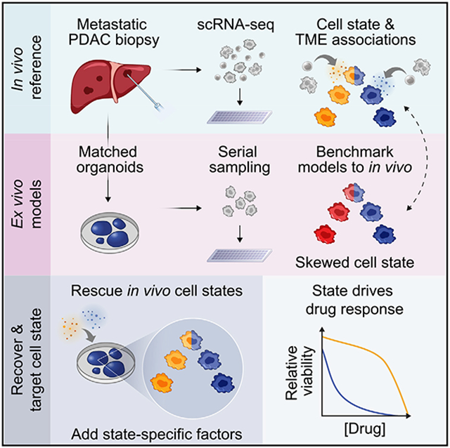
          Prognostically relevant RNA expression states exist in pancreatic ductal adenocarcinoma (PDAC), but our understanding of their drivers, stability, and relationship to therapeutic response is limited. To examine these attributes systematically, we profiled metastatic biopsies and matched organoid models at single-cell resolution. In vivo, we identify a new intermediate PDAC transcriptional cell state and uncover distinct site- and state-specific tumor microenvironments (TMEs). Benchmarking models against this reference map, we reveal strong culture-specific biases in cancer cell transcriptional state representation driven by altered TME signals. We restore expression state heterogeneity by adding back in vivo-relevant factors and show plasticity in culture models. Further, we prove that non-genetic modulation of cell state can strongly influence drug responses, uncovering state-specific vulnerabilities. This work provides a broadly applicable framework for aligning cell states across in vivo and ex vivo settings, identifying drivers of transcriptional plasticity and manipulating cell state to target associated vulnerabilities.

          Graphical Abstract

          In brief

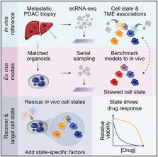
          Systematic profiling of metastatic pancreatic cancer biopsies and matched organoid models provides a view of cellular states, their regulation by the tumor microenvironment, and the ability to modulate these states to impact drug responses.

          Related collections

          Most cited references79

          • Record: found
          • Abstract: found
          • Article: not found
          Is Open Access

          The Cancer Genome Atlas Pan-Cancer analysis project.

          The Cancer Genome Atlas (TCGA) Research Network has profiled and analyzed large numbers of human tumors to discover molecular aberrations at the DNA, RNA, protein and epigenetic levels. The resulting rich data provide a major opportunity to develop an integrated picture of commonalities, differences and emergent themes across tumor lineages. The Pan-Cancer initiative compares the first 12 tumor types profiled by TCGA. Analysis of the molecular aberrations and their functional roles across tumor types will teach us how to extend therapies effective in one cancer type to others with a similar genomic profile.
            Bookmark
            • Record: found
            • Abstract: found
            • Article: not found

            Highly Parallel Genome-wide Expression Profiling of Individual Cells Using Nanoliter Droplets.

            Cells, the basic units of biological structure and function, vary broadly in type and state. Single-cell genomics can characterize cell identity and function, but limitations of ease and scale have prevented its broad application. Here we describe Drop-seq, a strategy for quickly profiling thousands of individual cells by separating them into nanoliter-sized aqueous droplets, associating a different barcode with each cell's RNAs, and sequencing them all together. Drop-seq analyzes mRNA transcripts from thousands of individual cells simultaneously while remembering transcripts' cell of origin. We analyzed transcriptomes from 44,808 mouse retinal cells and identified 39 transcriptionally distinct cell populations, creating a molecular atlas of gene expression for known retinal cell classes and novel candidate cell subtypes. Drop-seq will accelerate biological discovery by enabling routine transcriptional profiling at single-cell resolution. VIDEO ABSTRACT.
              Bookmark
              • Record: found
              • Abstract: found
              • Article: not found

              Dissecting the multicellular ecosystem of metastatic melanoma by single-cell RNA-seq.

              To explore the distinct genotypic and phenotypic states of melanoma tumors, we applied single-cell RNA sequencing (RNA-seq) to 4645 single cells isolated from 19 patients, profiling malignant, immune, stromal, and endothelial cells. Malignant cells within the same tumor displayed transcriptional heterogeneity associated with the cell cycle, spatial context, and a drug-resistance program. In particular, all tumors harbored malignant cells from two distinct transcriptional cell states, such that tumors characterized by high levels of the MITF transcription factor also contained cells with low MITF and elevated levels of the AXL kinase. Single-cell analyses suggested distinct tumor microenvironmental patterns, including cell-to-cell interactions. Analysis of tumor-infiltrating T cells revealed exhaustion programs, their connection to T cell activation and clonal expansion, and their variability across patients. Overall, we begin to unravel the cellular ecosystem of tumors and how single-cell genomics offers insights with implications for both targeted and immune therapies.
                Bookmark

                Author and article information

                Journal
                0413066
                2830
                Cell
                Cell
                Cell
                0092-8674
                1097-4172
                20 January 2022
                09 December 2021
                08 February 2022
                : 184
                : 25
                : 6119-6137.e26
                Affiliations
                [1 ]Department of Medical Oncology, Dana-Farber Cancer Institute, Boston, MA 02215, USA
                [2 ]Broad Institute of MIT and Harvard, Cambridge, MA 02142, USA
                [3 ]Harvard Medical School, Boston, MA 02115, USA
                [4 ]Department of Medicine, Brigham and Women’s Hospital, Boston, MA 02115, USA
                [5 ]Institute for Medical Engineering & Science, Massachusetts Institute of Technology, Cambridge, MA 02139, USA
                [6 ]Koch Institute for Integrative Cancer Research, Massachusetts Institute of Technology, Cambridge, MA 02139, USA
                [7 ]Department of Chemistry, Massachusetts Institute of Technology, Cambridge, MA 02139, USA
                [8 ]Center for Computational Molecular Biology, Brown University, Providence, RI 02912, USA
                [9 ]Division of Applied Mathematics, Brown University, Providence, RI 02912, USA
                [10 ]Department of Informatics and Analytics, Dana-Farber Cancer Institute, Boston, MA 02215, USA
                [11 ]Center for Cancer Genomics, Dana-Farber Cancer Institute, Boston, MA 02215, USA
                [12 ]Department of Oncologic Pathology, Dana-Farber Cancer Institute, Boston, MA 02215, USA
                [13 ]Massachusetts General Hospital Cancer Center, Boston, MA 02114, USA
                [14 ]Department of Surgery, Brigham and Women’s Hospital, Boston, MA 02115, USA
                [15 ]Department of Biostatistics, Brown University, Providence, RI 02912, USA
                [16 ]Microsoft Research New England, Cambridge, MA 02142, USA
                [17 ]Department of Biological Engineering, Massachusetts Institute of Technology, Cambridge, MA 02142, USA
                [18 ]Department of Pathology, Brigham and Women’s Hospital, Boston, MA 02115, USA
                [19 ]Ragon Institute of MGH, MIT, and Harvard, Cambridge, MA 02139, USA
                [20 ]These authors contributed equally
                [21 ]These authors contributed equally
                [22 ]Senior author
                [23 ]Lead contact
                Author notes

                AUTHOR CONTRIBUTIONS

                Conceptualization, S.R., P.S.W., B.M.W., W.C.H., A.J.A., A.K.S.; Methodology, S.R., P.S.W., A.W.N., H.L.W., L.C., J.A.N., B.M.W., W.C.H., A.J.A., A. K.S.; Validation, S.R., P.S.W., A.W.N., H.L.W., K.E.L., J.A.N., B.M.W., W.C.H., A.J.A., A.K.S.; Formal analysis, S.R., P.S.W., A.W.N., H.L.W., A.D., K.S.K., A.A.B., N.L., L.F.S., Y.Y.L., A.D. Cherniack, M.A.B., E.F.C., M.Y.T., J. M.M., L.C.; Investigation, S.R., P.S.W., A.W.N., H.L.W., K.E.L., J.G.-R., R.L.K., N.M., M.S.R., R.W.S.N., J.W., I.W.; Resources, S.A.V., A.D. Costa, E.K.H., D.Y.R., L.K.B., A.R., B.E.J., E.T.S., R.J.S., G.I.S, T.E.C., K.P., D.A.R., K.N., J.M.C., J.A.N., B.M.W.; Data Curation, P.S.W., S.R., A.W.N., H.L.W., K.S.K.; Writing – original draft, S.R., P.S.W., A.W.N., H.L.W.; Writing – review & editing, S.R., P.S.W., A.W.N., H.L.W., A.M.J., L.C., J.A.N., S.R.M., B.M.W., W.C.H., A.J.A., A.K.S.; Visualization, S.R., P.S.W., A.W.N., H.L.W.; Supervision, B.E.J., T.E.J., L.C., J.A.N., S.R.M., B.M.W., W.C.H., A.J.A., A.K.S.; Project administration, S.R., P.S.W., B.M.W., W.C.H., A.J.A., A.K.S.; Funding acquisition, S.R., P.S.W., S.R.M., B.M.W., W.C.H., A.J.A., A.K.S.

                Article
                NIHMS1758273
                10.1016/j.cell.2021.11.017
                8822455
                34890551
                5d84c0a2-591a-43b1-be71-9d54de91991c

                This is an open access article under the CC BY-NC-ND license ( http://creativecommons.org/licenses/by-nc-nd/4.0/).

                History
                Categories
                Article

                Cell biology
                Cell biology

                Comments

                Comment on this article