99
views
0
recommends
+1 Recommend
0 collections
    0
    shares
      • Record: found
      • Abstract: found
      • Article: found
      Is Open Access

      PINK1 phosphorylates ubiquitin to activate Parkin E3 ubiquitin ligase activity

      research-article

      Read this article at

      Bookmark
          There is no author summary for this article yet. Authors can add summaries to their articles on ScienceOpen to make them more accessible to a non-specialist audience.

          Abstract

          PINK1 phosphorylates ubiquitin, which then binds to Parkin and activates its E3 ligase activity, leading to induction of selective autophagy of damaged mitochondria.

          Abstract

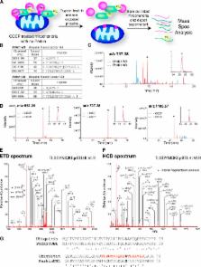
          PINK1 kinase activates the E3 ubiquitin ligase Parkin to induce selective autophagy of damaged mitochondria. However, it has been unclear how PINK1 activates and recruits Parkin to mitochondria. Although PINK1 phosphorylates Parkin, other PINK1 substrates appear to activate Parkin, as the mutation of all serine and threonine residues conserved between Drosophila and human, including Parkin S65, did not wholly impair Parkin translocation to mitochondria. Using mass spectrometry, we discovered that endogenous PINK1 phosphorylated ubiquitin at serine 65, homologous to the site phosphorylated by PINK1 in Parkin’s ubiquitin-like domain. Recombinant TcPINK1 directly phosphorylated ubiquitin and phospho-ubiquitin activated Parkin E3 ubiquitin ligase activity in cell-free assays. In cells, the phosphomimetic ubiquitin mutant S65D bound and activated Parkin. Furthermore, expression of ubiquitin S65A, a mutant that cannot be phosphorylated by PINK1, inhibited Parkin translocation to damaged mitochondria. These results explain a feed-forward mechanism of PINK1-mediated initiation of Parkin E3 ligase activity.

          Related collections

          Most cited references22

          • Record: found
          • Abstract: found
          • Article: not found

          Global survey of phosphotyrosine signaling identifies oncogenic kinases in lung cancer.

          Despite the success of tyrosine kinase-based cancer therapeutics, for most solid tumors the tyrosine kinases that drive disease remain unknown, limiting our ability to identify drug targets and predict response. Here we present the first large-scale survey of tyrosine kinase activity in lung cancer. Using a phosphoproteomic approach, we characterize tyrosine kinase signaling across 41 non-small cell lung cancer (NSCLC) cell lines and over 150 NSCLC tumors. Profiles of phosphotyrosine signaling are generated and analyzed to identify known oncogenic kinases such as EGFR and c-Met as well as novel ALK and ROS fusion proteins. Other activated tyrosine kinases such as PDGFRalpha and DDR1 not previously implicated in the genesis of NSCLC are also identified. By focusing on activated cell circuitry, the approach outlined here provides insight into cancer biology not available at the chromosomal and transcriptional levels and can be applied broadly across all human cancers.
            Bookmark
            • Record: found
            • Abstract: found
            • Article: found
            Is Open Access

            PINK1 is activated by mitochondrial membrane potential depolarization and stimulates Parkin E3 ligase activity by phosphorylating Serine 65

            Summary Missense mutations in PTEN-induced kinase 1 (PINK1) cause autosomal-recessive inherited Parkinson's disease (PD). We have exploited our recent discovery that recombinant insect PINK1 is catalytically active to test whether PINK1 directly phosphorylates 15 proteins encoded by PD-associated genes as well as proteins reported to bind PINK1. We have discovered that insect PINK1 efficiently phosphorylates only one of these proteins, namely the E3 ligase Parkin. We have mapped the phosphorylation site to a highly conserved residue within the Ubl domain of Parkin at Ser65. We show that human PINK1 is specifically activated by mitochondrial membrane potential (Δψm) depolarization, enabling it to phosphorylate Parkin at Ser65. We further show that phosphorylation of Parkin at Ser65 leads to marked activation of its E3 ligase activity that is prevented by mutation of Ser65 or inactivation of PINK1. We provide evidence that once activated, PINK1 autophosphorylates at several residues, including Thr257, which is accompanied by an electrophoretic mobility band-shift. These results provide the first evidence that PINK1 is activated following Δψm depolarization and suggest that PINK1 directly phosphorylates and activates Parkin. Our findings indicate that monitoring phosphorylation of Parkin at Ser65 and/or PINK1 at Thr257 represent the first biomarkers for examining activity of the PINK1-Parkin signalling pathway in vivo. Our findings also suggest that small molecule activators of Parkin that mimic the effect of PINK1 phosphorylation may confer therapeutic benefit for PD.
              Bookmark
              • Record: found
              • Abstract: found
              • Article: not found

              Phosphate-binding tag, a new tool to visualize phosphorylated proteins.

              We introduce two methods for the visualization of phosphorylated proteins using alkoxide-bridged dinuclear metal (i.e. Zn(2+) or Mn(2+)) complexes as novel phosphate-binding tag (Phos-tag) molecules. Both Zn(2+)- and Mn(2+)-Phos-tag molecules preferentially capture phosphomonoester dianions bound to Ser, Thr, and Tyr residues. One method is based on an ECL system using biotin-pendant Zn(2+)-Phos-tag and horseradish peroxidase-conjugated streptavidin. We demonstrate the electroblotting analyses of protein phosphorylation status by the phosphate-selective ECL signals. Another method is based on the mobility shift of phosphorylated proteins in SDS-PAGE with polyacrylamide-bound Mn(2+)-Phos-tag. Phosphorylated proteins in the gel are visualized as slower migration bands compared with corresponding dephosphorylated proteins. We demonstrate the kinase and phosphatase assays by phosphate affinity electrophoresis (Mn(2+)-Phos-tag SDS-PAGE).
                Bookmark

                Author and article information

                Journal
                J Cell Biol
                J. Cell Biol
                jcb
                jcb
                The Journal of Cell Biology
                The Rockefeller University Press
                0021-9525
                1540-8140
                28 April 2014
                : 205
                : 2
                : 143-153
                Affiliations
                [1 ]Biochemistry Section, Surgical Neurology Branch , and [2 ]Protein/Peptide Sequencing Facility, National Institute of Neurological Disorders and Stroke, National Institutes of Health, Bethesda, MD 20824
                Author notes
                Correspondence to Richard J. Youle: youler@ 123456ninds.nih.gov
                Article
                201402104
                10.1083/jcb.201402104
                4003245
                24751536
                5b719d70-2e7d-42cd-907e-64aae0856e43
                Copyright @ 2014

                This article is distributed under the terms of an Attribution–Noncommercial–Share Alike–No Mirror Sites license for the first six months after the publication date (see http://www.rupress.org/terms). After six months it is available under a Creative Commons License (Attribution–Noncommercial–Share Alike 3.0 Unported license, as described at http://creativecommons.org/licenses/by-nc-sa/3.0/).

                History
                : 20 February 2014
                : 2 April 2014
                Categories
                Research Articles
                Report

                Cell biology
                Cell biology

                Comments

                Comment on this article