Nanoscale Infrared Spectroscopy and Chemometrics Enable Detection of Intracellular Protein Distribution – ScienceOpen
33
views
0
recommends
+1 Recommend
0 collections
    0
    shares
      • Record: found
      • Abstract: found
      • Article: not found

      Nanoscale Infrared Spectroscopy and Chemometrics Enable Detection of Intracellular Protein Distribution

      research-article

      Read this article at

      ScienceOpenPublisherPMC
      Bookmark
          There is no author summary for this article yet. Authors can add summaries to their articles on ScienceOpen to make them more accessible to a non-specialist audience.

          Abstract

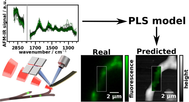
          Determination of the intracellular location of proteins is one of the fundamental tasks of microbiology. Conventionally, label-based microscopy and super-resolution techniques are employed. In this work, we demonstrate a new technique that can determine intracellular protein distribution at nanometer spatial resolution. This method combines nanoscale spatial resolution chemical imaging using the photothermal-induced resonance (PTIR) technique with multivariate modeling to reveal the intracellular distribution of cell components. Here, we demonstrate its viability by imaging the distribution of major cellulases and xylanases in Trichoderma reesei using the colocation of a fluorescent label (enhanced yellow fluorescence protein, EYFP) with the target enzymes to calibrate the chemometric model. The obtained partial least squares model successfully shows the distribution of these proteins inside the cell and opens the door for further studies on protein secretion mechanisms using PTIR.

          Related collections

          Most cited references78

          • Record: found
          • Abstract: found
          • Article: not found

          Infrared spectroscopy of proteins.

          This review discusses the application of infrared spectroscopy to the study of proteins. The focus is on the mid-infrared spectral region and the study of protein reactions by reaction-induced infrared difference spectroscopy.
            Bookmark
            • Record: found
            • Abstract: found
            • Article: not found

            Fourier transform infrared spectroscopic analysis of protein secondary structures.

            Infrared spectroscopy is one of the oldest and well established experimental techniques for the analysis of secondary structure of polypeptides and proteins. It is convenient, non-destructive, requires less sample preparation, and can be used under a wide variety of conditions. This review introduces the recent developments in Fourier transform infrared (FTIR) spectroscopy technique and its applications to protein structural studies. The experimental skills, data analysis, and correlations between the FTIR spectroscopic bands and protein secondary structure components are discussed. The applications of FTIR to the secondary structure analysis, conformational changes, structural dynamics and stability studies of proteins are also discussed.
              Bookmark
              • Record: found
              • Abstract: found
              • Article: not found

              Using Fourier transform IR spectroscopy to analyze biological materials.

              IR spectroscopy is an excellent method for biological analyses. It enables the nonperturbative, label-free extraction of biochemical information and images toward diagnosis and the assessment of cell functionality. Although not strictly microscopy in the conventional sense, it allows the construction of images of tissue or cell architecture by the passing of spectral data through a variety of computational algorithms. Because such images are constructed from fingerprint spectra, the notion is that they can be an objective reflection of the underlying health status of the analyzed sample. One of the major difficulties in the field has been determining a consensus on spectral pre-processing and data analysis. This manuscript brings together as coauthors some of the leaders in this field to allow the standardization of methods and procedures for adapting a multistage approach to a methodology that can be applied to a variety of cell biological questions or used within a clinical setting for disease screening or diagnosis. We describe a protocol for collecting IR spectra and images from biological samples (e.g., fixed cytology and tissue sections, live cells or biofluids) that assesses the instrumental options available, appropriate sample preparation, different sampling modes as well as important advances in spectral data acquisition. After acquisition, data processing consists of a sequence of steps including quality control, spectral pre-processing, feature extraction and classification of the supervised or unsupervised type. A typical experiment can be completed and analyzed within hours. Example results are presented on the use of IR spectra combined with multivariate data processing.
                Bookmark

                Author and article information

                Journal
                Anal Chem
                Anal Chem
                ac
                ancham
                Analytical Chemistry
                American Chemical Society
                0003-2700
                1520-6882
                01 December 2020
                15 December 2020
                : 92
                : 24
                : 15719-15725
                Affiliations
                []Institute of Chemical Technologies and Analytics, TU Wien , Vienna 1060, Austria
                []Institute of Chemical, Environmental and Bioscience Engineering, TU Wien , Vienna 1060, Austria
                Author notes
                Article
                10.1021/acs.analchem.0c02228
                7745202
                33259186
                5ad8a6a9-f57d-40b6-a84d-a13d8af4479f
                © 2020 American Chemical Society

                This is an open access article published under a Creative Commons Attribution (CC-BY) License, which permits unrestricted use, distribution and reproduction in any medium, provided the author and source are cited.

                History
                : 25 May 2020
                : 16 November 2020
                Categories
                Article
                Custom metadata
                ac0c02228
                ac0c02228

                Analytical chemistry
                Analytical chemistry

                Comments

                Comment on this article

                scite_
                14
                0
                18
                0
                Smart Citations
                14
                0
                18
                0
                Citing PublicationsSupportingMentioningContrasting
                View Citations

                See how this article has been cited at scite.ai

                scite shows how a scientific paper has been cited by providing the context of the citation, a classification describing whether it supports, mentions, or contrasts the cited claim, and a label indicating in which section the citation was made.

                Similar content93

                Cited by7

                Most referenced authors1,742