19
views
0
recommends
+1 Recommend
0 collections
    0
    shares
      • Record: found
      • Abstract: found
      • Article: not found

      An Objective Comparison of Cell Tracking Algorithms

      research-article
      1 , 1 , 2 , 3 , 4 , 5 , 1 , 1 , 1 , 6 , 6 , 5 , 2 , 7 , 8 , 8 , 9 , 10 , 11 , 12 , 13 , 13 , 14 , 14 , 3 , 3 , 15 , 15 , 4 , 4 , 16 , 17 , 16 , 18 , 18 , 19 , 19 , 6 , 20 , 21 , 1 , 22 , 23
      Nature methods

      Read this article at

      ScienceOpenPublisherPMC
      Bookmark
          There is no author summary for this article yet. Authors can add summaries to their articles on ScienceOpen to make them more accessible to a non-specialist audience.

          Abstract

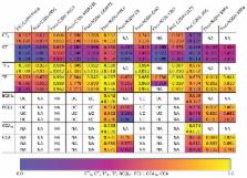
          We present a combined report on the results of three editions of the Cell Tracking Challenge, an ongoing initiative aimed at promoting the development and objective evaluation of cell tracking algorithms. With twenty-one participating algorithms and a data repository consisting of thirteen datasets of various microscopy modalities, the challenge displays today’s state of the art in the field. We analyze the results using performance measures for segmentation and tracking that rank all participating methods. We also analyze the performance of all algorithms in terms of biological measures and their practical usability. Even though some methods score high in all technical aspects, not a single one obtains fully correct solutions. We show that methods that either take prior information into account using learning strategies or analyze cells in a global spatio-temporal video context perform better than other methods under the segmentation and tracking scenarios included in the challenge.

          Related collections

          Most cited references41

          • Record: found
          • Abstract: found
          • Article: not found

          NIH Image to ImageJ: 25 years of image analysis.

          For the past 25 years NIH Image and ImageJ software have been pioneers as open tools for the analysis of scientific images. We discuss the origins, challenges and solutions of these two programs, and how their history can serve to advise and inform other software projects.
            Bookmark
            • Record: found
            • Abstract: found
            • Article: found
            Is Open Access

            U-Net: Convolutional Networks for Biomedical Image Segmentation

            There is large consent that successful training of deep networks requires many thousand annotated training samples. In this paper, we present a network and training strategy that relies on the strong use of data augmentation to use the available annotated samples more efficiently. The architecture consists of a contracting path to capture context and a symmetric expanding path that enables precise localization. We show that such a network can be trained end-to-end from very few images and outperforms the prior best method (a sliding-window convolutional network) on the ISBI challenge for segmentation of neuronal structures in electron microscopic stacks. Using the same network trained on transmitted light microscopy images (phase contrast and DIC) we won the ISBI cell tracking challenge 2015 in these categories by a large margin. Moreover, the network is fast. Segmentation of a 512x512 image takes less than a second on a recent GPU. The full implementation (based on Caffe) and the trained networks are available at http://lmb.informatik.uni-freiburg.de/people/ronneber/u-net .
              Bookmark
              • Record: found
              • Abstract: found
              • Article: found
              Is Open Access

              Objective comparison of particle tracking methods

              The first community competition designed to objectively compare the performance of particle tracking algorithms provides valuable practical information for both users and developers. Supplementary information The online version of this article (doi:10.1038/nmeth.2808) contains supplementary material, which is available to authorized users.
                Bookmark

                Author and article information

                Journal
                101215604
                32338
                Nat Methods
                Nat. Methods
                Nature methods
                1548-7091
                1548-7105
                6 October 2017
                30 October 2017
                December 2017
                30 April 2018
                : 14
                : 12
                : 1141-1152
                Affiliations
                [1 ]Centre for Biomedical Image Analysis, Masaryk University, Brno, Czech Republic
                [2 ]ACCESS Linnaeus Centre, KTH Royal Institute of Technology, Stockholm, Sweden
                [3 ]Computer Science Department and BIOSS Centre for Biological Signalling Studies University of Freiburg, Germany
                [4 ]Heidelberg Collaboratory for Image Processing, IWR, University of Heidelberg, Germany
                [5 ]Biomedical Computer Vision Group, Dept. Bioinformatics and Functional Genomics, BIOQUANT, IPMB, University of Heidelberg and DKFZ, Heidelberg, Germany
                [6 ]Biomedical Imaging Group Rotterdam, Departments of Medical Informatics and Radiology, Erasmus University Medical Center Rotterdam, Rotterdam, the Netherlands
                [7 ]Baxter Laboratory for Stem Cell Biology, Department of Microbiology and Immunology, and Institute for Stem Cell Biology and Regenerative Medicine, Stanford University School of Medicine, Stanford, CA, USA
                [8 ]Division of Image Processing, Department of Radiology, Leiden University Medical Center, Leiden, the Netherlands
                [9 ]Intelligent Systems Department, Delft University of Technology, Delft, the Netherlands
                [10 ]Institute of Molecular and Cell Biology, A*Star, Singapore
                [11 ]Department of Engineering, University of Nottingham, United Kingdom
                [12 ]Faculty of Engineering, University of Nottingham, Ningbo, China
                [13 ]BioImage Analysis Unit, Institut Pasteur, Paris, France
                [14 ]Research Centre in Biomedical Engineering, School of Mathematics, Computer Science and Engineering, City University of London, United Kingdom
                [15 ]Group for Automated Image and Data Analysis, Institute for Applied Computer Science, Karlsruhe Institute of Technology, Eggenstein-Leopoldshafen, Germany
                [16 ]i3S - Instituto de Investigação e Inovação em Saúde, Universidade do Porto, Porto, Portugal
                [17 ]Facultade de Engenharia, Universidade do Porto, Porto, Portugal
                [18 ]S3IT, University of Zurich, Switzerland
                [19 ]Max Planck Institute of Molecular Cell Biology and Genetics, Dresden, Germany
                [20 ]Bioengineering and Aerospace Engineering Department, Universidad Carlos III de Madrid, Spain
                [21 ]Instituto de Investigación Sanitaria Gregorio Marañon, Madrid, Spain
                [22 ]CIBERONC, IDISNA and Program of Solid Tumors and Biomarkers, Center for Applied Medical Research, University of Navarra, Pamplona, Spain
                [23 ]Bioengineering Department, TECNUN School of Engineering, University of Navarra, San Sebastián, Spain
                Author notes
                [* ]Corresponding author ( codesolorzano@ 123456unav.es )
                [a]

                Current affiliation: Max Planck Institute of Molecular Cell Biology and Genetics, Dresden, Germany

                [b]

                Current affiliation: DeepMind, London, UK

                [c]

                Current affiliation: Definiens AG, Munich, Germany

                [d]

                Current affiliation: National Heart Research Institute Singapore (NHRIS), National Heart Centre Singapore (NHCS), Singapore

                [e]

                Current affiliation: Computer Vision Institute, College of Computer Science and Software Engineering, Shenzhen University, Shenzhen, China

                [**]

                These authors contributed equally to this work

                Article
                NIHMS910062
                10.1038/nmeth.4473
                5777536
                29083403
                598226fb-b2c2-4563-bfc7-d64b0909d870

                Users may view, print, copy, and download text and data-mine the content in such documents, for the purposes of academic research, subject always to the full Conditions of use: http://www.nature.com/authors/editorial_policies/license.html#terms

                History
                Categories
                Article

                Life sciences
                Life sciences

                Comments

                Comment on this article