53
views
0
recommends
+1 Recommend
0 collections
    8
    shares
      • Record: found
      • Abstract: found
      • Article: found
      Is Open Access

      Promotion of allergic immune responses by intranasally-administrated nanosilica particles in mice

      research-article

      Read this article at

      Bookmark
          There is no author summary for this article yet. Authors can add summaries to their articles on ScienceOpen to make them more accessible to a non-specialist audience.

          Abstract

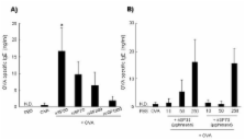
          With the increase in use of nanomaterials, there is growing concern regarding their potential health risks. However, few studies have assessed the role of the different physical characteristics of nanomaterials in allergic responses. Here, we examined whether intranasally administered silica particles of various sizes have the capacity to promote allergic immune responses in mice. We used nanosilica particles with diameters of 30 or 70 nm (nSP30 or nSP70, respectively), and conventional micro-sized silica particles with diameters of 300 or 1000 nm (nSP300 or mSP1000, respectively). Mice were intranasally exposed to ovalbumin (OVA) plus each silica particle, and the levels of OVA-specific antibodies (Abs) in the plasma were determined. Intranasal exposure to OVA plus smaller nanosilica particles tended to induce a higher level of OVA-specific immunoglobulin (Ig) E, IgG and IgG1 Abs than did exposure to OVA plus larger silica particles. Splenocytes from mice exposed to OVA plus nSP30 secreted higher levels of Th2-type cytokines than mice exposed to OVA alone. Taken together, these results indicate that nanosilica particles can induce allergen-specific Th2-type allergic immune responses in vivo. This study provides the foundations for the establishment of safe and effective forms of nanosilica particles.

          Related collections

          Most cited references18

          • Record: found
          • Abstract: found
          • Article: not found

          Size-dependent proinflammatory effects of ultrafine polystyrene particles: a role for surface area and oxidative stress in the enhanced activity of ultrafines.

          Studies into the effects of ultrafine particles in the lung have shown adverse effects considered to be due in part to the particle size. Air pollution particles (PM(10)) are associated with exacerbations of respiratory disease and deaths from cardiovascular causes in epidemiological studies and the ultrafine fraction of PM(10) has been hypothesized to play an important role. The aim of the present study was to investigate proinflammatory responses to various sizes of polystyrene particles as a simple model of particles of varying size including ultrafine. In the animal model, we demonstrated that there was a significantly greater neutrophil influx into the rat lung after instillation of 64-nm polystyrene particles compared with 202- and 535-nm particles and this was mirrored in other parameters of lung inflammation, such as increased protein and lactate dehydrogenase in bronchoalveolar lavage. When surface area instilled was plotted against inflammation, these two variables were directly proportional and the line passed through zero. This suggests that surface area drives inflammation in the short term and that ultrafine particles cause a greater inflammatory response because of the greater surface area they possess. In vitro, we measured the changes in intracellular calcium concentration in mono mac 6 cells in view of the potential role of calcium as a signaling molecule. Calcium changes after particle exposure may be important in leading to proinflammatory gene expression such as chemokines. We demonstrated that only ultrafine polystyrene particles induced a significant increase in cytosolic calcium ion concentration. Experiments using dichlorofluorescin diacetate demonstrated greater oxidant activity of the ultrafine particles, which may explain their activity in these assays. There were significant increases in IL-8 gene expression in A549 epithelial cells after treatment with the ultrafine particles but not particles of other sizes. These findings suggest that ultrafine particles composed of low-toxicity material such as polystyrene have proinflammatory activity as a consequence of their large surface area. This supports a role for such particles in the adverse health effects of PM(10). Copyright 2001 Academic Press.
            Bookmark
            • Record: found
            • Abstract: found
            • Article: not found

            Signaling by ROS drives inflammasome activation.

            Inflammasomes are innate immune signaling pathways that sense pathogens and injury to direct the proteolytic maturation of inflammatory cytokines such as IL-1beta and IL-18. Among inflammasomes, the NLRP3/NALP3 inflammasome is the most studied. However, little is known on the molecular mechanisms that mediate its assembly and activation. Recent findings suggest that ROS are produced by NLRP3/NALP3 activators and are essential secondary messengers signaling NLRP3/NALP3 inflammasome activation.
              Bookmark
              • Record: found
              • Abstract: found
              • Article: not found

              Organically modified silica nanoparticles: a nonviral vector for in vivo gene delivery and expression in the brain.

              This article reports on the application of organically modified silica (ORMOSIL) nanoparticles as a nonviral vector for efficient in vivo gene delivery. Highly monodispersed, stable aqueous suspension of nanoparticles, surface-functionalized with amino groups for binding of DNA, were prepared and characterized. Stereotaxic injections of nanoparticles, complexed with plasmid DNA encoding for EGFP, into the mouse ventral midbrain and into lateral ventricle, allowed us to fluorescently visualize the extensive transfection of neuronal-like cells in substantia nigra and areas surrounding the lateral ventricle. No ORMOSIL-based toxicity was observed 4 weeks after transfection. The efficiency of transfection equaled or exceeded that obtained in studies using a viral vector. An in vivo optical imaging technique (a fiber-based confocal fluorescent imaging system) provided an effective means to show the retention of viability of the transfected cells. The ORMOSIL-mediated transfections also were used to manipulate the biology of the neural stem/progenitor cells in vivo. Transfection of a plasmid expressing the nucleus-targeting fibroblast growth factor receptor type 1 resulted in significant inhibition of the in vivo incorporation of bromodeoxyuridine into the DNA of the cells in the subventricular zone and the adjacent rostral migratory stream. This in vivo approach shows that the nuclear receptor can control the proliferation of the stem/progenitor cells in this region of the brain. The results of this nanomedicine approach using ORMOSIL nanoparticles as a nonviral gene delivery platform have a promising future direction for effective therapeutic manipulation of the neural stem/progenitor cells as well as in vivo targeted brain therapy.
                Bookmark

                Author and article information

                Journal
                Nanoscale Res Lett
                Nanoscale Research Letters
                Springer
                1931-7573
                1556-276X
                2011
                4 March 2011
                : 6
                : 1
                : 195
                Affiliations
                [1 ]Department of Toxicology and Safety Science, Graduate School of Pharmaceutical Sciences, Osaka University, 1-6, Yamadaoka, Suita, Osaka 565-0871, Japan
                [2 ]Laboratory of Biopharmaceutical Research, National Institute of Biomedical Innovation, 7-6-8 Saito-asagi, Ibaraki, Osaka 567-0085, Japan
                [3 ]The Center for Advanced Medical Engineering and Informatics, Osaka University, 1-6, Yamadaoka, Suita, Osaka 565-0871, Japan
                [4 ]Department of Biomedical Innovation, Graduate school of Pharmaceutical Sciences, Osaka University, 7-6-8 Saito-asagi, Ibaraki, Osaka 567-0085, Japan
                Article
                1556-276X-6-195
                10.1186/1556-276X-6-195
                3211251
                21711705
                5963404a-a217-410b-841b-547fa496e850
                Copyright ©2011 Yoshida et al; licensee Springer.

                This is an Open Access article distributed under the terms of the Creative Commons Attribution License ( http://creativecommons.org/licenses/by/2.0), which permits unrestricted use, distribution, and reproduction in any medium, provided the original work is properly cited.

                History
                : 7 October 2010
                : 4 March 2011
                Categories
                Nano Express

                Nanomaterials
                Nanomaterials

                Comments

                Comment on this article