74
views
0
recommends
+1 Recommend
0 collections
    0
    shares
      • Record: found
      • Abstract: found
      • Article: found
      Is Open Access

      Impacted Mandibular Third Molars: Review of Literature and a Proposal of a Combined Clinical and Radiological Classification

      review-article

      Read this article at

      Bookmark
          There is no author summary for this article yet. Authors can add summaries to their articles on ScienceOpen to make them more accessible to a non-specialist audience.

          Abstract

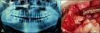
          Tooth impaction is a pathological situation where a tooth fails to attain its normal functional position. Impacted third molars are commonly encountered in routine dental practice. The impaction rate is higher for third molars when compared with other teeth. The mandibular third molar impaction is said to be due to the inadequate space between the distal of the second mandibular molar and the anterior border of the ascending ramus of the mandible. Impacted teeth may remain asymptomatic or may be associated with various pathologies such as caries, pericoronitis, cysts, tumors, and also root resorption of the adjacent tooth. Even though various classifications exist in the literature, none of those address the combined clinical and radiologic assessment of the impacted third molar. Literature search using the advanced features of various databases such as PubMed, Scopus, Embase, Google Scholar, Directory of Open Access Journals and Cochrane electronic databases was carried out. Keywords like impaction, mandibular third molar, impacted mandibular third molar, complications, anatomy, inferior alveolar nerve injury, lingual nerve injury were used to search the databases. A total of 826 articles were screened, and 50 articles were included in the review which was obtained from 1980 to February 2015. In the present paper, the authors have proposed a classification based on clinical and radiological assessment of the impacted mandibular third molar.

          Related collections

          Most cited references51

          • Record: found
          • Abstract: found
          • Article: not found

          Third molar outcomes from age 18 to 26: findings from a population-based New Zealand longitudinal study.

          The purpose of this study was to describe the presence and impaction status of people's third molars at age 18 years, as well as the observed changes in their clinical status between ages 18 and 26 years. Eight hundred twenty-one participants in a prospective cohort study were dentally examined at ages 18 and 26 years, and panoramic radiographs were taken at age 18 years but not at 26 years. For each tooth, its radiographic impaction status at age 18 years was compared with the clinical status by age 26 years. Of the 2857 third molars assessed at age 18 years, 92.8% were able to be followed up clinically at age 26 years. Approximately 54.9% of the teeth that were not impacted by age 18 had erupted by 26 years. Of the teeth that were impacted by age 18, 33.7% had fully erupted by age 26, 31.4% had been extracted, and 13.1% remained unerupted. Of the maxillary teeth that were categorized as "impacted" at age 18 years, 36.2% had fully erupted by age 26, whereas only 25.6% of the mandibular teeth had done so (P <.01). Fewer mandibular teeth than maxillary teeth remained unerupted by the time the patient was 26 years old (27.4% and 41.4%, respectively; P <.01), but there was no significant difference between the jaws in the proportion of impacted teeth at age 18 years that had been extracted by age 26 years (29.6% and 30.3%, respectively). For mesioangularly impacted third molars, 39.3% of maxillary teeth and 20.4% of mandibular teeth had fully erupted by age 26. Of the third molars that were mesioangularly impacted at age 18 years, 39.3% of maxillary teeth and 20.4% of mandibular teeth had fully erupted by age 26 years, whereas almost one-third of each had been extracted. Of the distoangularly impacted third molars, 20.4% of the maxillary teeth and one-third of the mandibular teeth had erupted by age 26, with 21.6% of the maxillary teeth and 31.6% of the mandibular teeth having been extracted. Other than horizontally impacted third molars, a substantial proportion of other impaction types do erupt fully, and radiographically apparent impaction in late adolescence should not be sufficient grounds for their prophylactic removal in the absence of other clinical indications.
            Bookmark
            • Record: found
            • Abstract: found
            • Article: found
            Is Open Access

            Mandibular Third Molar Impaction: Review of Literature and a Proposal of a Classification

            ABSTRACT Objectives The purpose of present article was to review impacted mandibular third molar aetiology, clinical anatomy, radiologic examination, surgical treatment and possible complications, as well as to create new mandibular third molar impaction and extraction difficulty degree classification based on anatomical and radiologic findings and literature review results. Material and Methods Literature was selected through a search of PubMed, Embase and Cochrane electronic databases. The keywords used for search were mandibular third molar, impacted mandibular third molar, inferior alveolar nerve injury third molar, lingual nerve injury third molar. The search was restricted to English language articles, published from 1976 to April 2013. Additionally, a manual search in the major anatomy and oral surgery journals and books was performed. The publications there selected by including clinical and human anatomy studies. Results In total 75 literature sources were obtained and reviewed. Impacted mandibular third molar aetiology, clinical anatomy, radiographic examination, surgical extraction of and possible complications, classifications and risk factors were discussed. New mandibular third molar impaction and extraction difficulty degree classification based on anatomical and radiologic findings and literature review results was proposed. Conclusions The classification proposed here based on anatomical and radiological impacted mandibular third molar features is promising to be a helpful tool for impacted tooth assessment as well as for planning for surgical operation. Further clinical studies should be conducted for new classification validation and reliability evaluation.
              Bookmark
              • Record: found
              • Abstract: found
              • Article: not found

              The incidence of unerupted permanent teeth and related clinical cases.

              The impacted or mal-erupted tooth and its associated pathosis provide great diagnostic challenges to the dentist. The frequency of occurrence of impaction was found to be over 10,000 in 5,000 Army recruits. The type and position of impaction are summarized in tables. The incidence of impactions was higher than previously reported and may reflect some sampling bias. The incidence of impacted second and fourth molars is higher than expected. Impactions or mal-eruptions were seen to involve every permanent tooth except the mandibular incisors and first molars. Impactions can occur because of malpositioning of the tooth bud or obstruction in the path of eruption. However, the exact mechanism is still unknown. Six unusual cases of impacted permanent teeth are described to illustrate the importance of panoramic radiographs in the discovery of some anomalously located impactions.
                Bookmark

                Author and article information

                Journal
                Ann Med Health Sci Res
                Ann Med Health Sci Res
                AMHSR
                Annals of Medical and Health Sciences Research
                Medknow Publications & Media Pvt Ltd (India )
                2141-9248
                2277-9205
                Jul-Aug 2015
                : 5
                : 4
                : 229-234
                Affiliations
                [1] Department of Oral Medicine and Radiology, College of Dentistry, Al Jouf University, Sakakah, Al Jouf, Kingdom of Saudi Arabia
                Author notes
                Address for correspondence: Dr. Santosh Patil, College of Dentistry, Al Jouf University, Sakakah, Al Jouf, Kingdom of Saudi Arabia. E-mail: drpsantosh@ 123456gmail.com
                Article
                AMHSR-5-229
                10.4103/2141-9248.160177
                4512113
                26229709
                58297d4f-44dc-475c-b38c-738522b15325
                Copyright: © Annals of Medical and Health Sciences Research

                This is an open-access article distributed under the terms of the Creative Commons Attribution-Noncommercial-Share Alike 3.0 Unported, which permits unrestricted use, distribution, and reproduction in any medium, provided the original work is properly cited.

                History
                Categories
                Review Article

                Medicine
                classification,complications,impacted tooth,management,third molar
                Medicine
                classification, complications, impacted tooth, management, third molar

                Comments

                Comment on this article