31
views
0
recommends
+1 Recommend
0 collections
    0
    shares
      • Record: found
      • Abstract: found
      • Article: found
      Is Open Access

      Musculoskeletal Geometry, Muscle Architecture and Functional Specialisations of the Mouse Hindlimb

      research-article

      Read this article at

      Bookmark
          There is no author summary for this article yet. Authors can add summaries to their articles on ScienceOpen to make them more accessible to a non-specialist audience.

          Abstract

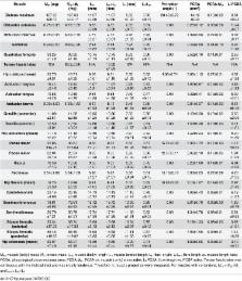
          Mice are one of the most commonly used laboratory animals, with an extensive array of disease models in existence, including for many neuromuscular diseases. The hindlimb is of particular interest due to several close muscle analogues/homologues to humans and other species. A detailed anatomical study describing the adult morphology is lacking, however. This study describes in detail the musculoskeletal geometry and skeletal muscle architecture of the mouse hindlimb and pelvis, determining the extent to which the muscles are adapted for their function, as inferred from their architecture. Using I 2KI enhanced microCT scanning and digital segmentation, it was possible to identify 39 distinct muscles of the hindlimb and pelvis belonging to nine functional groups. The architecture of each of these muscles was determined through microdissections, revealing strong architectural specialisations between the functional groups. The hip extensors and hip adductors showed significantly stronger adaptations towards high contraction velocities and joint control relative to the distal functional groups, which exhibited larger physiological cross sectional areas and longer tendons, adaptations for high force output and elastic energy savings. These results suggest that a proximo-distal gradient in muscle architecture exists in the mouse hindlimb. Such a gradient has been purported to function in aiding locomotor stability and efficiency. The data presented here will be especially valuable to any research with a focus on the architecture or gross anatomy of the mouse hindlimb and pelvis musculature, but also of use to anyone interested in the functional significance of muscle design in relation to quadrupedal locomotion.

          Related collections

          Most cited references46

          • Record: found
          • Abstract: found
          • Article: not found

          The placental mammal ancestor and the post-K-Pg radiation of placentals.

          To discover interordinal relationships of living and fossil placental mammals and the time of origin of placentals relative to the Cretaceous-Paleogene (K-Pg) boundary, we scored 4541 phenomic characters de novo for 86 fossil and living species. Combining these data with molecular sequences, we obtained a phylogenetic tree that, when calibrated with fossils, shows that crown clade Placentalia and placental orders originated after the K-Pg boundary. Many nodes discovered using molecular data are upheld, but phenomic signals overturn molecular signals to show Sundatheria (Dermoptera + Scandentia) as the sister taxon of Primates, a close link between Proboscidea (elephants) and Sirenia (sea cows), and the monophyly of echolocating Chiroptera (bats). Our tree suggests that Placentalia first split into Xenarthra and Epitheria; extinct New World species are the oldest members of Afrotheria.
            Bookmark
            • Record: found
            • Abstract: found
            • Article: not found

            Scaling body support in mammals: limb posture and muscle mechanics.

            The scaling of bone and muscle geometry in mammals suggests that peak stresses (ratio of force to cross-sectional area) acting in these two support elements increase with increasing body size. Observations of stresses acting in the limb bones of different sized mammals during strenuous activity, however, indicate that peak bone stress is independent of size (maintaining a safety factor of between 2 and 4). It appears that similar peak bone stresses and muscle stresses in large and small mammals are achieved primarily by a size-dependent change in locomotor limb posture: small animals run with crouched postures, whereas larger species run more upright. By adopting an upright posture, large animals align their limbs more closely with the ground reaction force, substantially reducing the forces that their muscles must exert (proportional to body mass) and hence, the forces that their bones must resist, to counteract joint moments. This change in limb posture to maintain locomotor stresses within safe limits, however, likely limits the maneuverability and accelerative capability of large animals.
              Bookmark
              • Record: found
              • Abstract: found
              • Article: not found

              Functional and clinical significance of skeletal muscle architecture.

              Skeletal muscle architecture is the structural property of whole muscles that dominates their function. This review describes the basic architectural properties of human upper and lower extremity muscles. The designs of various muscle groups in humans and other species are analyzed from the point of view of optimizing function. Muscle fiber arrangement and motor unit arrangement is discussed in terms of the control of movement. Finally, the ability of muscles to change their architecture in response to immobilization, eccentric exercise, and surgical tendon transfer is reviewed. Future integrative physiological studies will provide insights into the mechanisms by which such adaptations occur. It is likely that muscle fibers transduce both stress and strain and respond by modifying sarcomere number in a way more suited to the new biomechanical environment. Copyright 2000 John Wiley & Sons, Inc.
                Bookmark

                Author and article information

                Contributors
                Role: Editor
                Journal
                PLoS One
                PLoS ONE
                plos
                plosone
                PLoS ONE
                Public Library of Science (San Francisco, CA USA )
                1932-6203
                26 April 2016
                2016
                : 11
                : 4
                : e0147669
                Affiliations
                [1 ]Neuromuscular Diseases Group, Comparative Biomedical Sciences, Royal Veterinary College, 4 Royal College Street, London, NW1 0TU, United Kingdom
                [2 ]Structure and Motion Lab, Comparative Biomedical Sciences, Royal Veterinary College, Hawkshead Lane, Hatfield, Hertfordshire, AL9 7TA, United Kingdom
                [3 ]Department of Bioengineering, College of Engineering, Temple University, 1947 N. 12th Street, Philadelphia, PA 19122, United States of America
                University of Sydney, AUSTRALIA
                Author notes

                Competing Interests: The authors have declared that no competing interests exist.

                Conceived and designed the experiments: JRH AJS DJW JPC. Performed the experiments: JPC OC. Analyzed the data: JPC. Contributed reagents/materials/analysis tools: JPC OC DJW. Wrote the paper: JPC OC AJS JRH DJW.

                Article
                PONE-D-15-45553
                10.1371/journal.pone.0147669
                4846001
                27115354
                581017ce-7a42-4508-8529-66ceaf71ad64
                © 2016 Charles et al

                This is an open access article distributed under the terms of the Creative Commons Attribution License, which permits unrestricted use, distribution, and reproduction in any medium, provided the original author and source are credited.

                History
                : 29 October 2015
                : 6 January 2016
                Page count
                Figures: 3, Tables: 7, Pages: 21
                Funding
                Funded by: funder-id http://dx.doi.org/10.13039/501100000268, Biotechnology and Biological Sciences Research Council;
                Award ID: BB/J021504/1
                Award Recipient :
                Funded by: funder-id http://dx.doi.org/10.13039/100006754, U.S. Army Research Laboratory;
                Award ID: 64929EG
                Award Recipient :
                Funding provided by Biotechnology and Biological Sciences Research Council ( http://www.bbsrc.ac.uk/) BB/J021504/1 and US Army Research Office ( http://www.arl.army.mil/) 64929EG. The funders had no role in study design, data collection and analysis, decision to publish, or preparation of the manuscript.
                Categories
                Research Article
                Biology and Life Sciences
                Anatomy
                Musculoskeletal System
                Limbs (Anatomy)
                Legs
                Ankles
                Medicine and Health Sciences
                Anatomy
                Musculoskeletal System
                Limbs (Anatomy)
                Legs
                Ankles
                Biology and Life Sciences
                Physiology
                Muscle Physiology
                Muscle Functions
                Medicine and Health Sciences
                Physiology
                Muscle Physiology
                Muscle Functions
                Biology and Life Sciences
                Anatomy
                Musculoskeletal System
                Limbs (Anatomy)
                Legs
                Knees
                Medicine and Health Sciences
                Anatomy
                Musculoskeletal System
                Limbs (Anatomy)
                Legs
                Knees
                Biology and Life Sciences
                Anatomy
                Musculoskeletal System
                Pelvis
                Hip
                Medicine and Health Sciences
                Anatomy
                Musculoskeletal System
                Pelvis
                Hip
                Biology and Life Sciences
                Anatomy
                Biological Tissue
                Connective Tissue
                Tendons
                Medicine and Health Sciences
                Anatomy
                Biological Tissue
                Connective Tissue
                Tendons
                Biology and Life Sciences
                Organisms
                Animals
                Vertebrates
                Amniotes
                Mammals
                Rodents
                Mice
                Biology and Life Sciences
                Cell Biology
                Cellular Types
                Animal Cells
                Muscle Fibers
                Biology and Life Sciences
                Anatomy
                Musculoskeletal System
                Muscles
                Muscle Fibers
                Medicine and Health Sciences
                Anatomy
                Musculoskeletal System
                Muscles
                Muscle Fibers
                Biology and Life Sciences
                Biomechanics
                Biological Locomotion
                Biology and Life Sciences
                Physiology
                Biological Locomotion
                Medicine and Health Sciences
                Physiology
                Biological Locomotion
                Custom metadata
                All relevant data are within the paper and its Supporting Information files.

                Uncategorized
                Uncategorized

                Comments

                Comment on this article