14
views
0
recommends
+1 Recommend
1 collections
    0
    shares
      • Record: found
      • Abstract: found
      • Article: found
      Is Open Access

      An alpaca nanobody neutralizes SARS-CoV-2 by blocking receptor interaction

      research-article

      Read this article at

      Bookmark
          There is no author summary for this article yet. Authors can add summaries to their articles on ScienceOpen to make them more accessible to a non-specialist audience.

          Abstract

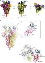
          SARS-CoV-2 enters host cells through an interaction between the spike glycoprotein and the angiotensin converting enzyme 2 (ACE2) receptor. Directly preventing this interaction presents an attractive possibility for suppressing SARS-CoV-2 replication. Here, we report the isolation and characterization of an alpaca-derived single domain antibody fragment, Ty1, that specifically targets the receptor binding domain (RBD) of the SARS-CoV-2 spike, directly preventing ACE2 engagement. Ty1 binds the RBD with high affinity, occluding ACE2. A cryo-electron microscopy structure of the bound complex at 2.9 Å resolution reveals that Ty1 binds to an epitope on the RBD accessible in both the ‘up’ and ‘down’ conformations, sterically hindering RBD-ACE2 binding. While fusion to an Fc domain renders Ty1 extremely potent, Ty1 neutralizes SARS-CoV-2 spike pseudovirus as a 12.8 kDa nanobody, which can be expressed in high quantities in bacteria, presenting opportunities for manufacturing at scale. Ty1 is therefore an excellent candidate as an intervention against COVID-19.

          Abstract

          Here, Hanke et al. immunize an alpaca with SARS-CoV-2 spike protein domains and identify a nanobody that binds the receptor binding domain of spike in both the up and down conformations and sterically hinders ACE2 engagement.

          Related collections

          Most cited references25

          • Record: found
          • Abstract: found
          • Article: found
          Is Open Access

          Cryo-EM structure of the 2019-nCoV spike in the prefusion conformation

          Structure of the nCoV trimeric spike The World Health Organization has declared the outbreak of a novel coronavirus (2019-nCoV) to be a public health emergency of international concern. The virus binds to host cells through its trimeric spike glycoprotein, making this protein a key target for potential therapies and diagnostics. Wrapp et al. determined a 3.5-angstrom-resolution structure of the 2019-nCoV trimeric spike protein by cryo–electron microscopy. Using biophysical assays, the authors show that this protein binds at least 10 times more tightly than the corresponding spike protein of severe acute respiratory syndrome (SARS)–CoV to their common host cell receptor. They also tested three antibodies known to bind to the SARS-CoV spike protein but did not detect binding to the 2019-nCoV spike protein. These studies provide valuable information to guide the development of medical counter-measures for 2019-nCoV. Science, this issue p. 1260
            Bookmark
            • Record: found
            • Abstract: found
            • Article: not found

            Structure, Function, and Antigenicity of the SARS-CoV-2 Spike Glycoprotein

            Summary The emergence of SARS-CoV-2 has resulted in >90,000 infections and >3,000 deaths. Coronavirus spike (S) glycoproteins promote entry into cells and are the main target of antibodies. We show that SARS-CoV-2 S uses ACE2 to enter cells and that the receptor-binding domains of SARS-CoV-2 S and SARS-CoV S bind with similar affinities to human ACE2, correlating with the efficient spread of SARS-CoV-2 among humans. We found that the SARS-CoV-2 S glycoprotein harbors a furin cleavage site at the boundary between the S1/S2 subunits, which is processed during biogenesis and sets this virus apart from SARS-CoV and SARS-related CoVs. We determined cryo-EM structures of the SARS-CoV-2 S ectodomain trimer, providing a blueprint for the design of vaccines and inhibitors of viral entry. Finally, we demonstrate that SARS-CoV S murine polyclonal antibodies potently inhibited SARS-CoV-2 S mediated entry into cells, indicating that cross-neutralizing antibodies targeting conserved S epitopes can be elicited upon vaccination.
              Bookmark
              • Record: found
              • Abstract: found
              • Article: found
              Is Open Access

              Cell entry mechanisms of SARS-CoV-2

              Significance A key to curbing SARS-CoV-2 is to understand how it enters cells. SARS-CoV-2 and SARS-CoV both use human ACE2 as entry receptor and human proteases as entry activators. Using biochemical and pseudovirus entry assays and SARS-CoV as a comparison, we have identified key cell entry mechanisms of SARS-CoV-2 that potentially contribute to the immune evasion, cell infectivity, and wide spread of the virus. This study also clarifies conflicting reports from recent studies on cell entry of SARS-CoV-2. Finally, by highlighting the potency and the evasiveness of SARS-CoV-2, the study provides insight into intervention strategies that target its cell entry mechanisms.
                Bookmark

                Author and article information

                Contributors
                martin.hallberg@ki.se
                benjamin.murrell@ki.se
                gerald.mcinerney@ki.se
                Journal
                Nat Commun
                Nat Commun
                Nature Communications
                Nature Publishing Group UK (London )
                2041-1723
                4 September 2020
                4 September 2020
                2020
                : 11
                : 4420
                Affiliations
                [1 ]GRID grid.4714.6, ISNI 0000 0004 1937 0626, Department of Microbiology, Tumor and Cell Biology, , Karolinska Institutet, ; Stockholm, Sweden
                [2 ]GRID grid.7836.a, ISNI 0000 0004 1937 1151, Division of Virology, Institute of Infectious Diseases and Molecular Medicine, Faculty of Health Sciences, , University of Cape Town, ; Cape Town, South Africa
                [3 ]GRID grid.4714.6, ISNI 0000 0004 1937 0626, Department of Cell and Molecular Biology, , Karolinska Institutet, ; Stockholm, Sweden
                [4 ]GRID grid.24381.3c, ISNI 0000 0000 9241 5705, Science for Life Laboratory, Department of Medicine Solna, Karolinska Institutet, and Division of Infectious Diseases, , Karolinska University Hospital, ; Solna, Stockholm, Sweden
                [5 ]Karolinska Institutet VR-RÅC, Centre for Structural Systems Biology, Notkestraße 85, 22607 Hamburg, Germany
                Author information
                http://orcid.org/0000-0001-5514-2418
                http://orcid.org/0000-0003-4283-812X
                http://orcid.org/0000-0002-6781-0345
                http://orcid.org/0000-0002-0393-4445
                http://orcid.org/0000-0003-2257-7241
                Article
                18174
                10.1038/s41467-020-18174-5
                7473855
                32887876
                55e5990b-6ea0-4728-83b6-fc5d6cb2f88c
                © The Author(s) 2020

                Open Access This article is licensed under a Creative Commons Attribution 4.0 International License, which permits use, sharing, adaptation, distribution and reproduction in any medium or format, as long as you give appropriate credit to the original author(s) and the source, provide a link to the Creative Commons license, and indicate if changes were made. The images or other third party material in this article are included in the article’s Creative Commons license, unless indicated otherwise in a credit line to the material. If material is not included in the article’s Creative Commons license and your intended use is not permitted by statutory regulation or exceeds the permitted use, you will need to obtain permission directly from the copyright holder. To view a copy of this license, visit http://creativecommons.org/licenses/by/4.0/.

                History
                : 19 June 2020
                : 8 August 2020
                Categories
                Article
                Custom metadata
                © The Author(s) 2020

                Uncategorized
                antibodies,sars-cov-2,cryoelectron microscopy
                Uncategorized
                antibodies, sars-cov-2, cryoelectron microscopy

                Comments

                Comment on this article