30
views
0
recommends
+1 Recommend
0 collections
    0
    shares
      • Record: found
      • Abstract: found
      • Article: found
      Is Open Access

      MoVrp1, a putative verprolin protein, is required for asexual development and infection in the rice blast fungus Magnaporthe oryzae

      research-article

      Read this article at

      Bookmark
          There is no author summary for this article yet. Authors can add summaries to their articles on ScienceOpen to make them more accessible to a non-specialist audience.

          Abstract

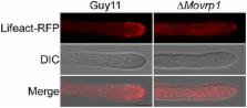
          Endocytosis is a crucial cellular process in eukaryotic cells which involves clathrin and/or adaptor proteins, lipid kinases, phosphatases and the actin cytoskeleton. Verprolin proteins, such as Vrp1 in Saccharomyces cerevisiae, are conserved family proteins that regulate actin binding and endocytosis. Here, we identified and characterized MoVrp1 as the yeast Vrp1 homolog in Magnaporthe oryzae. Deletion of the MoVRP1 gene resulted in defects in vegetative growth, asexual development, and infection of the host plant. The ∆Movrp1 mutants also exhibited decreased extracellular peroxidase and laccase activities and showed defects in colony pigmentation, hyphal surface hydrophobicity, cell wall integrity, autophagy, endocytosis, and secretion of avirulent effector. Our studies provided new evidences that MoVrp1 involved in actin cytoskeleton is important for growth, morphogenesis, cellular trafficking, and fungal pathogenesis.

          Related collections

          Most cited references63

          • Record: found
          • Abstract: found
          • Article: not found

          A new vital stain for visualizing vacuolar membrane dynamics and endocytosis in yeast

          SD Emr (1995)
          We have used a lipophilic styryl dye, N-(3-triethylammoniumpropyl)-4- (p-diethylaminophenyl-hexatrienyl) pyridinium dibromide (FM 4-64), as a vital stain to follow bulk membrane-internalization and transport to the vacuole in yeast. After treatment for 60 min at 30 degrees C, FM 4- 64 stained the vacuole membrane (ring staining pattern). FM 4-64 did not appear to reach the vacuole by passive diffusion because at 0 degree C it exclusively stained the plasma membrane (PM). The PM staining decreased after warming cells to 25 degrees C and small punctate structures became apparent in the cytoplasm within 5-10 min. After an additional 20-40 min, the PM and cytoplasmic punctate staining disappeared concomitant with staining of the vacuolar membrane. Under steady state conditions, FM 4-64 staining was specific for vacuolar membranes; other membrane structures were not stained. The dye served as a sensitive reporter of vacuolar dynamics, detecting such events as segregation structure formation during mitosis, vacuole fission/fusion events, and vacuolar morphology in different classes of vacuolar protein sorting (vps) mutants. A particularly striking pattern was observed in class E mutants (e.g., vps27) where 500-700 nm organelles (presumptive prevacuolar compartments) were intensely stained with FM 4- 64 while the vacuole membrane was weakly fluorescent. Internalization of FM 4-64 at 15 degrees C delayed vacuolar labeling and trapped FM 4- 64 in cytoplasmic intermediates between the PM and the vacuole. The intermediate structures in the cytoplasm are likely to be endosomes as their staining was temperature, time, and energy dependent. Interestingly, unlike Lucifer yellow uptake, vacuolar labeling by FM 4- 64 was not blocked in sec18, sec14, end3, and end4 mutants, but was blocked in sec1 mutant cells. Finally, using permeabilized yeast spheroplasts to reconstitute FM 4-64 transport, we found that delivery of FM 4-64 from the endosome-like intermediate compartment (labeled at 15 degrees C) to the vacuole was ATP and cytosol dependent. Thus, we show that FM 4-64 is a new vital stain for the vacuolar membrane, a marker for endocytic intermediates, and a fluor for detecting endosome to vacuole membrane transport in vitro.
            Bookmark
            • Record: found
            • Abstract: found
            • Article: not found

            Metabolism and functions of gamma-aminobutyric acid.

            B. Shelp (1999)
            Gamma-aminobutyric acid (GABA), a four-carbon non-protein amino acid, is a significant component of the free amino acid pool in most prokaryotic and eukaryotic organisms. In plants, stress initiates a signal-transduction pathway, in which increased cytosolic Ca2+ activates Ca2+/calmodulin-dependent glutamate decarboxylase activity and GABA synthesis. Elevated H+ and substrate levels can also stimulate glutamate decarboxylase activity. GABA accumulation probably is mediated primarily by glutamate decarboxylase. However, more information is needed concerning the control of the catabolic mitochondrial enzymes (GABA transaminase and succinic semialdehyde dehydrogenase) and the intracellular and intercellular transport of GABA. Experimental evidence supports the involvement of GABA synthesis in pH regulation, nitrogen storage, plant development and defence, as well as a compatible osmolyte and an alternative pathway for glutamate utilization. There is a need to identify the genes of enzymes involved in GABA metabolism, and to generate mutants with which to elucidate the physiological function(s) of GABA in plants.
              Bookmark
              • Record: found
              • Abstract: found
              • Article: not found

              Translocation of Magnaporthe oryzae effectors into rice cells and their subsequent cell-to-cell movement.

              Knowledge remains limited about how fungal pathogens that colonize living plant cells translocate effector proteins inside host cells to regulate cellular processes and neutralize defense responses. To cause the globally important rice blast disease, specialized invasive hyphae (IH) invade successive living rice (Oryza sativa) cells while enclosed in host-derived extrainvasive hyphal membrane. Using live-cell imaging, we identified a highly localized structure, the biotrophic interfacial complex (BIC), which accumulates fluorescently labeled effectors secreted by IH. In each newly entered rice cell, effectors were first secreted into BICs at the tips of the initially filamentous hyphae in the cell. These tip BICs were left behind beside the first-differentiated bulbous IH cells as the fungus continued to colonize the host cell. Fluorescence recovery after photobleaching experiments showed that the effector protein PWL2 (for prevents pathogenicity toward weeping lovegrass [Eragrostis curvula]) continued to accumulate in BICs after IH were growing elsewhere. PWL2 and BAS1 (for biotrophy-associated secreted protein 1), BIC-localized secreted proteins, were translocated into the rice cytoplasm. By contrast, BAS4, which uniformly outlines the IH, was not translocated into the host cytoplasm. Fluorescent PWL2 and BAS1 proteins that reached the rice cytoplasm moved into uninvaded neighbors, presumably preparing host cells before invasion. We report robust assays for elucidating the molecular mechanisms that underpin effector secretion into BICs, translocation to the rice cytoplasm, and cell-to-cell movement in rice.
                Bookmark

                Author and article information

                Journal
                Sci Rep
                Sci Rep
                Scientific Reports
                Nature Publishing Group
                2045-2322
                24 January 2017
                2017
                : 7
                : 41148
                Affiliations
                [1 ]Department of Plant Pathology, College of Plant Protection, Nanjing Agricultural University, and Key Laboratory of Integrated Management of Crop Diseases and Pests, Ministry of Education , Nanjing 210095, China
                [2 ]College of Forestry and Co-Innovation Center for Sustainable Forestry in Southern China, Nanjing Forestry University , Nanjing, Jiangsu 210037, China
                [3 ]Department of Pediatrics, Louisiana State University Health Sciences Center, New Orleans , Louisiana 70118, USA
                Author notes
                Article
                srep41148
                10.1038/srep41148
                5259722
                53ed2e5e-367d-462a-abe2-789889c6b830
                Copyright © 2017, The Author(s)

                This work is licensed under a Creative Commons Attribution 4.0 International License. The images or other third party material in this article are included in the article’s Creative Commons license, unless indicated otherwise in the credit line; if the material is not included under the Creative Commons license, users will need to obtain permission from the license holder to reproduce the material. To view a copy of this license, visit http://creativecommons.org/licenses/by/4.0/

                History
                : 28 September 2016
                : 15 December 2016
                Categories
                Article

                Uncategorized
                Uncategorized

                Comments

                Comment on this article