13
views
0
recommends
+1 Recommend
0 collections
    0
    shares
      • Record: found
      • Abstract: found
      • Article: found
      Is Open Access

      Predicting survival time of lung cancer patients using radiomic analysis

      research-article

      Read this article at

      Bookmark
          There is no author summary for this article yet. Authors can add summaries to their articles on ScienceOpen to make them more accessible to a non-specialist audience.

          Abstract

          Objectives

          This study investigates the prediction of Non-small cell lung cancer (NSCLC) patient survival outcomes based on radiomic texture and shape features automatically extracted from tumor image data.

          Materials and Methods

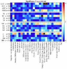
          Retrospective analysis involves CT scans of 315 NSCLC patients from The Cancer Imaging Archive (TCIA). A total of 24 image features are computed from labeled tumor volumes of patients within groups defined using NSCLC subtype and TNM staging information. Spearman’s rank correlation, Kaplan-Meier estimation and log-rank tests were used to identify features related to long/short NSCLC patient survival groups. Automatic random forest classification was used to predict patient survival group from multivariate feature data. Significance is assessed at P < 0.05 following Holm-Bonferroni correction for multiple comparisons.

          Results

          Significant correlations between radiomic features and survival were observed for four clinical groups: (group, [absolute correlation range]): (large cell carcinoma (LCC) [0.35, 0.43]), (tumor size T2, [0.31, 0.39]), (non lymph node metastasis N0, [0.3, 0.33]), (TNM stage I, [0.39, 0.48]). Significant log-rank relationships between features and survival time were observed for three clinical groups: (group, hazard ratio): (LCC, 3.0), (LCC, 3.9), (T2, 2.5) and (stage I, 2.9). Automatic survival prediction performance (i.e. below/above median) is superior for combined radiomic features with age-TNM in comparison to standard TNM clinical staging information (clinical group, mean area-under-the-ROC-curve (AUC)): (LCC, 75.73%), (N0, 70.33%), (T2, 70.28%) and (TNM-I, 76.17%).

          Conclusion

          Quantitative lung CT imaging features can be used as indicators of survival, in particular for patients with large-cell-carcinoma (LCC), primary-tumor-sizes (T2) and no lymph-node-metastasis (N0).

          Related collections

          Most cited references33

          • Record: found
          • Abstract: found
          • Article: not found

          The meaning and use of the area under a receiver operating characteristic (ROC) curve.

          A representation and interpretation of the area under a receiver operating characteristic (ROC) curve obtained by the "rating" method, or by mathematical predictions based on patient characteristics, is presented. It is shown that in such a setting the area represents the probability that a randomly chosen diseased subject is (correctly) rated or ranked with greater suspicion than a randomly chosen non-diseased subject. Moreover, this probability of a correct ranking is the same quantity that is estimated by the already well-studied nonparametric Wilcoxon statistic. These two relationships are exploited to (a) provide rapid closed-form expressions for the approximate magnitude of the sampling variability, i.e., standard error that one uses to accompany the area under a smoothed ROC curve, (b) guide in determining the size of the sample required to provide a sufficiently reliable estimate of this area, and (c) determine how large sample sizes should be to ensure that one can statistically detect differences in the accuracy of diagnostic techniques.
            Bookmark
            • Record: found
            • Abstract: found
            • Article: found
            Is Open Access

            Machine Learning methods for Quantitative Radiomic Biomarkers

            Radiomics extracts and mines large number of medical imaging features quantifying tumor phenotypic characteristics. Highly accurate and reliable machine-learning approaches can drive the success of radiomic applications in clinical care. In this radiomic study, fourteen feature selection methods and twelve classification methods were examined in terms of their performance and stability for predicting overall survival. A total of 440 radiomic features were extracted from pre-treatment computed tomography (CT) images of 464 lung cancer patients. To ensure the unbiased evaluation of different machine-learning methods, publicly available implementations along with reported parameter configurations were used. Furthermore, we used two independent radiomic cohorts for training (n = 310 patients) and validation (n = 154 patients). We identified that Wilcoxon test based feature selection method WLCX (stability = 0.84 ± 0.05, AUC = 0.65 ± 0.02) and a classification method random forest RF (RSD = 3.52%, AUC = 0.66 ± 0.03) had highest prognostic performance with high stability against data perturbation. Our variability analysis indicated that the choice of classification method is the most dominant source of performance variation (34.21% of total variance). Identification of optimal machine-learning methods for radiomic applications is a crucial step towards stable and clinically relevant radiomic biomarkers, providing a non-invasive way of quantifying and monitoring tumor-phenotypic characteristics in clinical practice.
              Bookmark
              • Record: found
              • Abstract: found
              • Article: found
              Is Open Access

              Predicting non-small cell lung cancer prognosis by fully automated microscopic pathology image features

              Lung cancer is the most prevalent cancer worldwide, and histopathological assessment is indispensable for its diagnosis. However, human evaluation of pathology slides cannot accurately predict patients' prognoses. In this study, we obtain 2,186 haematoxylin and eosin stained histopathology whole-slide images of lung adenocarcinoma and squamous cell carcinoma patients from The Cancer Genome Atlas (TCGA), and 294 additional images from Stanford Tissue Microarray (TMA) Database. We extract 9,879 quantitative image features and use regularized machine-learning methods to select the top features and to distinguish shorter-term survivors from longer-term survivors with stage I adenocarcinoma (P<0.003) or squamous cell carcinoma (P=0.023) in the TCGA data set. We validate the survival prediction framework with the TMA cohort (P<0.036 for both tumour types). Our results suggest that automatically derived image features can predict the prognosis of lung cancer patients and thereby contribute to precision oncology. Our methods are extensible to histopathology images of other organs.
                Bookmark

                Author and article information

                Journal
                Oncotarget
                Oncotarget
                Oncotarget
                ImpactJ
                Oncotarget
                Impact Journals LLC
                1949-2553
                28 November 2017
                1 November 2017
                : 8
                : 61
                : 104393-104407
                Affiliations
                1 Division of Radiation Oncology, McGill University, Montréal, Canada
                2 The Laboratory for Imagery, Vision and Artificial Intelligence, Ecole de Technologie Supérieure, Montréal, Canada
                Author notes
                Correspondence to: Ahmad Chaddad, ahmad.chaddad@ 123456mail.mcgill.ca
                Article
                22251
                10.18632/oncotarget.22251
                5732814
                29262648
                53a754b4-6d21-4820-8ae3-8e7fc6ddc96f
                Copyright: © 2017 Chaddad et al.

                This is an open-access article distributed under the terms of the Creative Commons Attribution License 3.0 (CC BY 3.0), which permits unrestricted use, distribution, and reproduction in any medium, provided the original author and source are credited.

                History
                : 30 May 2017
                : 2 October 2017
                Categories
                Research Paper

                Oncology & Radiotherapy
                lung cancer,nsclc,cancer staging,radiomics,texture features
                Oncology & Radiotherapy
                lung cancer, nsclc, cancer staging, radiomics, texture features

                Comments

                Comment on this article