97
views
0
recommends
+1 Recommend
0 collections
    0
    shares
      • Record: found
      • Abstract: found
      • Article: found
      Is Open Access

      Neural traces of stress: cortisol related sustained enhancement of amygdala-hippocampal functional connectivity

      research-article

      Read this article at

      Bookmark
          There is no author summary for this article yet. Authors can add summaries to their articles on ScienceOpen to make them more accessible to a non-specialist audience.

          Abstract

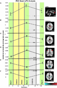
          Stressful experiences modulate neuro-circuitry function, and the temporal trajectory of these alterations, elapsing from early disturbances to late recovery, heavily influences resilience and vulnerability to stress. Such effects of stress may depend on processes that are engaged during resting-state, through active recollection of past experiences and anticipation of future events, all known to involve the default mode network (DMN). By inducing social stress and acquiring resting-state functional magnetic resonance imaging (fMRI) before stress, immediately following it, and 2 h later, we expanded the time-window for examining the trajectory of the stress response. Throughout the study repeated cortisol samplings and self-reports of stress levels were obtained from 51 healthy young males. Post-stress alterations were investigated by whole brain resting-state functional connectivity (rsFC) of two central hubs of the DMN: the posterior cingulate cortex (PCC) and hippocampus. Results indicate a ’recovery’ pattern of DMN connectivity, in which all alterations, ascribed to the intervening stress, returned to pre-stress levels. The only exception to this pattern was a stress-induced rise in amygdala-hippocampal connectivity, which was sustained for as long as 2 h following stress induction. Furthermore, this sustained enhancement of limbic connectivity was inversely correlated to individual stress-induced cortisol responsiveness (AUCi) and characterized only the group lacking such increased cortisol (i.e., non-responders). Our observations provide evidence of a prolonged post-stress response profile, characterized by both the comprehensive balance of most DMN functional connections and the distinct time and cortisol dependent ascent of intra-limbic connectivity. These novel insights into neuro-endocrine relations are another milestone in the ongoing search for individual markers in stress-related psychopathologies.

          Related collections

          Most cited references30

          • Record: found
          • Abstract: found
          • Article: not found

          Spontaneous low-frequency BOLD signal fluctuations: an fMRI investigation of the resting-state default mode of brain function hypothesis.

          Recent neuroimaging studies have lead to the proposal that rest is characterized by an organized, baseline level of activity, a default mode of brain function that is suspended during specific goal-oriented mental activity. Previous studies have shown that the primary function subserved by the default mode is that of an introspectively oriented, self-referential mode of mental activity. The default mode of brain function hypothesis is readdressed from the perspective of the presence of low-frequency blood oxygenation level-dependent (BOLD) functional magnetic resonance imaging (fMRI) signal changes (0.012-0.1 Hz) in the resting brain. The results show that the brain during rest is not tonically active in a single mode of brain function. Rather, the findings presented here suggest that the brain recurrently toggles between an introspectively oriented mode (default mode) and a state-of-mind that tentatively might be interpreted as an extrospectively oriented mode that involves a readiness and alertness to changes in the external and internal environment.
            Bookmark
            • Record: found
            • Abstract: found
            • Article: not found

            Functional connectivity of default mode network components: correlation, anticorrelation, and causality.

            The default mode network (DMN), based in ventromedial prefrontal cortex (vmPFC) and posterior cingulate cortex (PCC), exhibits higher metabolic activity at rest than during performance of externally oriented cognitive tasks. Recent studies have suggested that competitive relationships between the DMN and various task-positive networks involved in task performance are intrinsically represented in the brain in the form of strong negative correlations (anticorrelations) between spontaneous fluctuations in these networks. Most neuroimaging studies characterize the DMN as a homogenous network, thus few have examined the differential contributions of DMN components to such competitive relationships. Here, we examined functional differentiation within the DMN, with an emphasis on understanding competitive relationships between this and other networks. We used a seed correlation approach on resting-state data to assess differences in functional connectivity between these two regions and their anticorrelated networks. While the positively correlated networks for the vmPFC and PCC seeds largely overlapped, the anticorrelated networks for each showed striking differences. Activity in vmPFC negatively predicted activity in parietal visual spatial and temporal attention networks, whereas activity in PCC negatively predicted activity in prefrontal-based motor control circuits. Granger causality analyses suggest that vmPFC and PCC exert greater influence on their anticorrelated networks than the other way around, suggesting that these two default mode nodes may directly modulate activity in task-positive networks. Thus, the two major nodes comprising the DMN are differentiated with respect to the specific brain systems with which they interact, suggesting greater heterogeneity within this network than is commonly appreciated.
              Bookmark
              • Record: found
              • Abstract: found
              • Article: not found

              Response variation following trauma: a translational neuroscience approach to understanding PTSD.

              Exposure to traumatic stress is a requirement for the development of posttraumatic stress disorder (PTSD). However, because the majority of trauma-exposed persons do not develop PTSD, examination of the typical effects of a stressor will not identify the critical components of PTSD risk or pathogenesis. Rather, PTSD represents a specific phenotype associated with a failure to recover from the normal effects of trauma. Thus, research must focus on identifying pre- and posttraumatic risk factors that explain the development of the disorder and the failure to reinstate physiological homeostasis. In this review, we summarize what is known about the clinical and biological characteristics of PTSD and articulate some of the gaps in knowledge that can be addressed by basic neuroscience research. We emphasize how knowledge about individual differences related to genetic and epigenetic factors in behavioral and brain responses to stress offers the hope of a deeper understanding of PTSD.
                Bookmark

                Author and article information

                Journal
                Front Hum Neurosci
                Front Hum Neurosci
                Front. Hum. Neurosci.
                Frontiers in Human Neuroscience
                Frontiers Media S.A.
                1662-5161
                05 July 2013
                2013
                : 7
                : 313
                Affiliations
                [1] 1Functional Brain Center, Wohl Institute for Advanced Imaging, Sourasky Medical Center Tel-Aviv, Israel
                [2] 2Sackler Faculty of Medicine, Tel Aviv University Tel Aviv, Israel
                [3] 3Sagol School of Neuroscience, Tel Aviv University Tel Aviv, Israel
                [4] 4School of Psychological Sciences, Tel Aviv University Tel Aviv, Israel
                [5] 5Center for Depression, Anxiety, and Stress Research, Harvard Medical School, McLean Hospital Belmont, MA, USA
                [6] 6Sourasky Medical Center, Institute of Endocrinology, Metabolism, and Hypertension Tel Aviv, Israel
                [7] 7Division of Mental Health, Medical Corps, IDF Tel Hashomer, Israel
                [8] 8Section on Development and Affective Neuroscience of the National Institute of Mental Health, National Institutes of Health Bethesda, MD, USA
                [9] 9School of Education, Tel Aviv University Tel Aviv, Israel
                Author notes

                Edited by: Hadas Okon-Singer, University of Haifa, Israel

                Reviewed by: Judy A. Kipping, Max Planck Institute for Human Cognitive and Brain Sciences, Germany; Markus Muehlhan, Technische Universitaet Dresden, Germany

                *Correspondence: Talma Hendler, Wohl Institute for Advanced Imaging, Tel-Aviv Sourasky Medical Center, 6 Weizmann Street, Tel-Aviv 64239, Israel e-mail: talma@ 123456tasmc.health.gov.il;
                Sharon Vaisvaser, Functional Brain Center, Wohl Institute for Advanced Imaging, Sourasky Medical Center, Tel-Aviv, Israel e-mail: ysharonv@ 123456gmail.com
                Article
                10.3389/fnhum.2013.00313
                3701866
                23847492
                533d04a3-6940-49de-8248-9c53ba86311a
                Copyright © 2013 Vaisvaser, Lin, Admon, Podlipsky, Greenman, Stern, Fruchter, Wald, Pine, Tarrasch, Bar-Haim and Hendler.

                This is an open-access article distributed under the terms of the Creative Commons Attribution License, which permits use, distribution and reproduction in other forums, provided the original authors and source are credited and subject to any copyright notices concerning any third-party graphics etc.

                History
                : 15 January 2013
                : 10 June 2013
                Page count
                Figures: 5, Tables: 2, Equations: 0, References: 52, Pages: 11, Words: 7931
                Categories
                Neuroscience
                Original Research Article

                Neurosciences
                fmri,resting-state functional connectivity,default-mode network,recovery,limbic connectivity

                Comments

                Comment on this article