2
views
0
recommends
+1 Recommend
0 collections
    0
    shares
      • Record: found
      • Abstract: found
      • Article: found
      Is Open Access

      Altered hippocampus and amygdala subregion connectome hierarchy in major depressive disorder

      research-article

      Read this article at

      Bookmark
          There is no author summary for this article yet. Authors can add summaries to their articles on ScienceOpen to make them more accessible to a non-specialist audience.

          Abstract

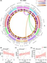
          The hippocampus and amygdala limbic structures are critical to the etiology of major depressive disorder (MDD). However, there are no high-resolution characterizations of the role of their subregions in the whole brain network (connectome). Connectomic examination of these subregions can uncover disorder-related patterns that are otherwise missed when treated as single structures. 38 MDD patients and 40 healthy controls (HC) underwent anatomical and diffusion imaging using 7-Tesla MRI. Whole-brain segmentation was performed along with hippocampus and amygdala subregion segmentation, each representing a node in the connectome. Graph theory analysis was applied to examine the importance of the limbic subregions within the brain network using centrality features measured by node strength (sum of weights of the node’s connections), Betweenness (number of shortest paths that traverse the node), and clustering coefficient (how connected the node’s neighbors are to one another and forming a cluster). Compared to HC, MDD patients showed decreased node strength of the right hippocampus cornu ammonis (CA) 3/4, indicating decreased connectivity to the rest of the brain, and decreased clustering coefficient of the right dentate gyrus, implying it is less embedded in a cluster. Additionally, within the MDD group, the greater the embedding of the right amygdala central nucleus (CeA) in a cluster, the greater the severity of depressive symptoms. The altered role of these limbic subregions in the whole-brain connectome is related to diagnosis and depression severity, contributing to our understanding of the limbic system involvement in MDD and may elucidate the underlying mechanisms of depression.

          Related collections

          Most cited references76

          • Record: found
          • Abstract: not found
          • Article: not found

          Controlling the False Discovery Rate: A Practical and Powerful Approach to Multiple Testing

            Bookmark
            • Record: found
            • Abstract: found
            • Article: not found

            G*Power 3: A flexible statistical power analysis program for the social, behavioral, and biomedical sciences

            G*Power (Erdfelder, Faul, & Buchner, 1996) was designed as a general stand-alone power analysis program for statistical tests commonly used in social and behavioral research. G*Power 3 is a major extension of, and improvement over, the previous versions. It runs on widely used computer platforms (i.e., Windows XP, Windows Vista, and Mac OS X 10.4) and covers many different statistical tests of the t, F, and chi2 test families. In addition, it includes power analyses for z tests and some exact tests. G*Power 3 provides improved effect size calculators and graphic options, supports both distribution-based and design-based input modes, and offers all types of power analyses in which users might be interested. Like its predecessors, G*Power 3 is free.
              Bookmark
              • Record: found
              • Abstract: found
              • Article: not found

              An automated labeling system for subdividing the human cerebral cortex on MRI scans into gyral based regions of interest.

              In this study, we have assessed the validity and reliability of an automated labeling system that we have developed for subdividing the human cerebral cortex on magnetic resonance images into gyral based regions of interest (ROIs). Using a dataset of 40 MRI scans we manually identified 34 cortical ROIs in each of the individual hemispheres. This information was then encoded in the form of an atlas that was utilized to automatically label ROIs. To examine the validity, as well as the intra- and inter-rater reliability of the automated system, we used both intraclass correlation coefficients (ICC), and a new method known as mean distance maps, to assess the degree of mismatch between the manual and the automated sets of ROIs. When compared with the manual ROIs, the automated ROIs were highly accurate, with an average ICC of 0.835 across all of the ROIs, and a mean distance error of less than 1 mm. Intra- and inter-rater comparisons yielded little to no difference between the sets of ROIs. These findings suggest that the automated method we have developed for subdividing the human cerebral cortex into standard gyral-based neuroanatomical regions is both anatomically valid and reliable. This method may be useful for both morphometric and functional studies of the cerebral cortex as well as for clinical investigations aimed at tracking the evolution of disease-induced changes over time, including clinical trials in which MRI-based measures are used to examine response to treatment.
                Bookmark

                Author and article information

                Contributors
                yael.jacob@mssm.edu
                Journal
                Transl Psychiatry
                Transl Psychiatry
                Translational Psychiatry
                Nature Publishing Group UK (London )
                2158-3188
                19 May 2022
                19 May 2022
                2022
                : 12
                : 209
                Affiliations
                [1 ]GRID grid.59734.3c, ISNI 0000 0001 0670 2351, Depression and Anxiety Center for Discovery and Treatment, Department of Psychiatry, , Icahn School of Medicine at Mount Sinai, ; New York, NY USA
                [2 ]GRID grid.59734.3c, ISNI 0000 0001 0670 2351, BioMedical Engineering and Imaging Institute, , Icahn School of Medicine at Mount Sinai, ; New York, NY USA
                [3 ]GRID grid.59734.3c, ISNI 0000 0001 0670 2351, Department of Psychiatry, , Icahn School of Medicine at Mount Sinai, ; New York, NY USA
                [4 ]GRID grid.59734.3c, ISNI 0000 0001 0670 2351, Department of Diagnostic, Molecular and Interventional Radiology, , Icahn School of Medicine at Mount Sinai, ; New York, NY USA
                [5 ]GRID grid.59734.3c, ISNI 0000 0001 0670 2351, Department of Neuroscience, , Icahn School of Medicine at Mount Sinai, ; New York, NY USA
                Author information
                http://orcid.org/0000-0003-3278-9264
                http://orcid.org/0000-0002-5862-6650
                http://orcid.org/0000-0001-6286-1242
                Article
                1976
                10.1038/s41398-022-01976-0
                9120054
                35589678
                f6a7c7fc-a6d5-4fae-97a1-ebe516e6ba2b
                © The Author(s) 2022

                Open Access This article is licensed under a Creative Commons Attribution 4.0 International License, which permits use, sharing, adaptation, distribution and reproduction in any medium or format, as long as you give appropriate credit to the original author(s) and the source, provide a link to the Creative Commons license, and indicate if changes were made. The images or other third party material in this article are included in the article’s Creative Commons license, unless indicated otherwise in a credit line to the material. If material is not included in the article’s Creative Commons license and your intended use is not permitted by statutory regulation or exceeds the permitted use, you will need to obtain permission directly from the copyright holder. To view a copy of this license, visit http://creativecommons.org/licenses/by/4.0/.

                History
                : 24 January 2022
                : 4 May 2022
                : 10 May 2022
                Funding
                Funded by: FundRef https://doi.org/10.13039/100000025, U.S. Department of Health & Human Services | NIH | National Institute of Mental Health (NIMH);
                Award ID: R01 MH109544
                Award ID: R01MH109544
                Award Recipient :
                Funded by: U.S. Department of Health & Human Services | NIH | National Institute of Mental Health (NIMH)
                Categories
                Article
                Custom metadata
                © The Author(s) 2022

                Clinical Psychology & Psychiatry
                neuroscience,depression
                Clinical Psychology & Psychiatry
                neuroscience, depression

                Comments

                Comment on this article