42
views
0
recommends
+1 Recommend
0 collections
    0
    shares
      • Record: found
      • Abstract: found
      • Article: found
      Is Open Access

      Total Flavonoids from Clinopodium chinense (Benth.) O. Ktze Protect against Doxorubicin-Induced Cardiotoxicity In Vitro and In Vivo

      research-article

      Read this article at

      Bookmark
          There is no author summary for this article yet. Authors can add summaries to their articles on ScienceOpen to make them more accessible to a non-specialist audience.

          Abstract

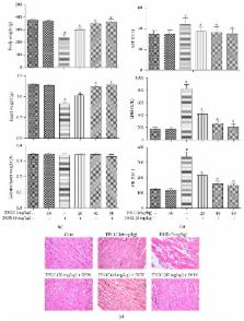
          Doxorubicin has cardiotoxic effects that limit its clinical benefit in cancer patients. This study aims to investigate the protective effects of the total flavonoids from Clinopodium chinense (Benth.) O. Ktze (TFCC) against doxorubicin- (DOX-) induced cardiotoxicity. Male rats were intraperitoneally injected with a single dose of DOX (3 mg/kg) every 2 days for three injections. Heart samples were collected 2 weeks after the last DOX dose and then analyzed. DOX delayed body and heart growth and caused cardiac tissue injury, oxidative stress, apoptotic damage, mitochondrial dysfunction, and Bcl-2 expression disturbance. Similar experiments in H9C2 cardiomyocytes showed that doxorubicin reduced cell viability, increased ROS generation and DNA fragmentation, disrupted mitochondrial membrane potential, and induced apoptotic cell death. However, TFCC pretreatment suppressed all of these adverse effects of doxorubicin. Signal transduction studies indicated that TFCC suppressed DOX-induced overexpression of p53 and phosphorylation of JNK, p38, and ERK. Studies with LY294002 (a PI3K/AKT inhibitor) demonstrated that the mechanism of TFCC-induced cardioprotection also involves activation of PI3K/AKT. These findings indicated the potential clinical application of TFCC in preventing DOX-induced cardiac oxidative stress.

          Related collections

          Most cited references50

          • Record: found
          • Abstract: found
          • Article: not found

          Taurine suppresses doxorubicin-triggered oxidative stress and cardiac apoptosis in rat via up-regulation of PI3-K/Akt and inhibition of p53, p38-JNK.

          The objective of the present study was to investigate the signaling mechanisms involved in the beneficial role of taurine against doxorubicin-induced cardiac oxidative stress. Male rats were administered doxorubicin. Hearts were collected 3 weeks after the last dose of doxorubicin and were analyzed. Doxorubicin administration retarded the growth of the body and the heart and caused injury in the cardiac tissue because of increased oxidative stress. Similar experiments with doxorubicin showed reduced cell viability, increased ROS generation, intracellular Ca(2+) and DNA fragmentation, disrupted mitochondrial membrane potential and apoptotic cell death in primary cultured neonatal rat cardiomyocytes. Signal transduction studies showed that doxorubicin increased p53, JNK, p38 and NFκB phosphorylation; decreased the levels of phospho ERK and Akt; disturbed the Bcl-2 family protein balance; activated caspase 12, caspase 9 and caspase 3; and induced cleavage of the PARP protein. However, taurine treatment or cardiomyocyte incubation with taurine suppressed all of the adverse effects of doxorubicin. Studies with several inhibitors, including PS-1145 (an IKK inhibitor), SP600125 (a JNK inhibitor), SB203580 (a p38 inhibitor) and LY294002 (a PI3-K/Akt inhibitor), demonstrated that the mechanism of taurine-induced cardio protection involves activation of specific survival signals and PI3-K/Akt as well as the inhibition of p53, JNK, p38 and NFκB. These novel findings suggest that taurine might have clinical implications for the prevention of doxorubicin-induced cardiac oxidative stress. Copyright © 2011 Elsevier Inc. All rights reserved.
            Bookmark
            • Record: found
            • Abstract: found
            • Article: not found

            The protective role of arjunolic acid against doxorubicin induced intracellular ROS dependent JNK-p38 and p53-mediated cardiac apoptosis.

            In spite of tremendous demand for the development and implementation of effective therapeutic strategies, limitations are still associated with doxorubicin-induced cardiotoxicity. Arjunolic acid (AA) has been shown to possess a multitude of biological functions. The purpose of the present study was to explore whether AA plays any protective role against doxorubicin-induced cardiotoxicity; and if so, what molecular mechanism it utilizes for its protective action. In rat cardiomyocytes, doxorubicin administration activated the proapoptotic p53, p38 and JNK MAPKs, Bax translocation, disrupted mitochondrial membrane potential, precipitated mitochondrion mediated caspase-dependent apoptotic signalling and reduced viability of cardiomyocytes. Doxorubicin exposure increases dichlorofluorescein (DCF) intensity corresponding to the intracellular H(2)O(2) generation in myocytes; catalase (CAT) treatment, however, reduced this intensity and preserves cell viability. Intracellular H(2)O(2) thus produced now activates the p38-JNK and p53-mediated pathways. CAT treatment also markedly decreased the doxorubicin-mediated activation of p38 and JNK, suggesting that H(2)O(2) is involved in the activation of MAPKs. Blockage of p53 and p38-JNK by pharmacological inhibitors also suppressed the doxorubicin-induced apoptosis with the concomitant inhibition of anti-apoptotic Bcl-2 family proteins. AA treatment ameliorates nearly all of these apoptotic actions of doxorubicin and preserves cell viability. Similarly, rats treated with doxorubicin displayed retarded growth of body and heart as well as elevated apoptotic indices in heart tissue, whereas AA treatment effectively neutralised all these doxorubicin-induced cardiac-abnormalities. Combining all, our results suggest that doxorubicin induces cardiac apoptosis via the activation of JNK-p38 and p53-mediated signalling pathways, where H(2)O(2) acts as the mediators of these pathways. AA can effectively and extensively counteract this action of doxorubicin, and may potentially protect the heart and cardiomyocytes from the severe doxorubicin-induced cardiovascular burden. Copyright © 2011 Elsevier Ltd. All rights reserved.
              Bookmark
              • Record: found
              • Abstract: found
              • Article: not found

              Mitochondrial proliferation during apoptosis induced by anticancer agents: effects of doxorubicin and mitoxantrone on cancer and cardiac cells.

              Doxorubicin is one of the most largely prescribed antitumor drug for the treatment of breast, liver and colon cancers as well as leukemia, but the cardiotoxicity of this anthracycline derivative limits its clinical use. Although doxorubicin is toxic to both cancer and cardiac cells, there are evidences suggesting that the mechanism of cell death is different for the two cell types. To investigate further this issue, we have compared the proapoptotic effects of doxorubicin and the functionally related anthracenedione compound mitoxantrone, which is also used in the clinic for the treatment of cancer. After evaluating the toxicity of the two drugs to mammary adenocarcinoma MTLn3 cells and H9C2 cardiomyocytes, we dissected the drug-induced apoptotic machinery by measuring the effects on the cell cycle progression, DNA condensation and fragmentation, production of endogenous peroxides and caspase activation. Both doxorubicin and mitoxantrone are potent inducers of apoptosis in H9C2 cardiomyocytes and MTLn3 breast cancer cells, but there are significant differences between the two cell types in terms of kinetics and order of the events. In particular, flow cytometry measurements of drug-induced changes in mitochondrial transmembrane potential and mitochondrial mass with different fluorescent probes suggested that the two drugs induced a progressive increase in mitochondrial mass in the cancer cells but not in the cardiac cells. The hypothesis was validated by means of electron microscopy, which revealed a significant increase in the number of mitochondria in drug-treated MTLn3 but not in H9C2 cells. The mitochondrial proliferation precedes the nuclear apoptosis in doxorubicin-treated MTLn3 cells. The changes in the architecture and number of mitochondria are linked to the drug-induced perturbation of the cell cycle progression and apoptosis. The proliferation of mitochondria could explain the higher toxicity of doxorubicin to cancer cells compared to cardiac cells and this suggests novel therapeutic opportunities to better control the cardiotoxicity of anthracyclines.
                Bookmark

                Author and article information

                Journal
                Evid Based Complement Alternat Med
                Evid Based Complement Alternat Med
                ECAM
                Evidence-based Complementary and Alternative Medicine : eCAM
                Hindawi Publishing Corporation
                1741-427X
                1741-4288
                2015
                16 February 2015
                16 February 2015
                : 2015
                : 472565
                Affiliations
                1Institute of Medicinal Plant Development, Chinese Academy of Medical Science, Peking Union Medical College, No. 151, North Road Malianwa, Haidian District, Beijing 100094, China
                2Academy of Forestry, Baishan, Jilin 134302, China
                3Academy of Chinese Materia Medica, Wenzhou Medical College, Wenzhou, Zhejiang 325035, China
                4Harbin University of Commerce, Xuehai Street, Songbei District, Harbin, Heilongjiang 150028, China
                5Department of Pharmacology, Heilongjiang University of Chinese Medicine, Harbin, Heilongjiang 150040, China
                Author notes
                *Gui Bo Sun: 97095112@ 123456qq.com and

                Academic Editor: Shuang-En Chuang

                Article
                10.1155/2015/472565
                4346128
                25784945
                511dc437-57da-475b-88ca-dee3ed72cb7a
                Copyright © 2015 Rong Chang Chen et al.

                This is an open access article distributed under the Creative Commons Attribution License, which permits unrestricted use, distribution, and reproduction in any medium, provided the original work is properly cited.

                History
                : 7 September 2014
                : 4 December 2014
                : 22 December 2014
                Categories
                Research Article

                Complementary & Alternative medicine
                Complementary & Alternative medicine

                Comments

                Comment on this article