0
views
0
recommends
+1 Recommend
0 collections
    0
    shares
      • Record: found
      • Abstract: found
      • Article: found
      Is Open Access

      Exploring deep learning radiomics for classifying osteoporotic vertebral fractures in X-ray images

      research-article

      Read this article at

      Bookmark
          There is no author summary for this article yet. Authors can add summaries to their articles on ScienceOpen to make them more accessible to a non-specialist audience.

          Abstract

          Purpose

          To develop and validate a deep learning radiomics (DLR) model that uses X-ray images to predict the classification of osteoporotic vertebral fractures (OVFs).

          Material and methods

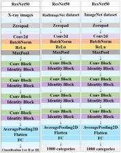
          The study encompassed a cohort of 942 patients, involving examinations of 1076 vertebrae through X-ray, CT, and MRI across three distinct hospitals. The OVFs were categorized as class 0, 1, or 2 based on the Assessment System of Thoracolumbar Osteoporotic Fracture. The dataset was divided randomly into four distinct subsets: a training set comprising 712 samples, an internal validation set with 178 samples, an external validation set containing 111 samples, and a prospective validation set consisting of 75 samples. The ResNet-50 architectural model was used to implement deep transfer learning (DTL), undergoing -pre-training separately on the RadImageNet and ImageNet datasets. Features from DTL and radiomics were extracted and integrated using X-ray images. The optimal fusion feature model was identified through least absolute shrinkage and selection operator logistic regression. Evaluation of the predictive capabilities for OVFs classification involved eight machine learning models, assessed through receiver operating characteristic curves employing the “One-vs-Rest” strategy. The Delong test was applied to compare the predictive performance of the superior RadImageNet model against the ImageNet model.

          Results

          Following pre-training separately on RadImageNet and ImageNet datasets, feature selection and fusion yielded 17 and 12 fusion features, respectively. Logistic regression emerged as the optimal machine learning algorithm for both DLR models. Across the training set, internal validation set, external validation set, and prospective validation set, the macro-average Area Under the Curve (AUC) based on the RadImageNet dataset surpassed those based on the ImageNet dataset, with statistically significant differences observed (P<0.05). Utilizing the binary “One-vs-Rest” strategy, the model based on the RadImageNet dataset demonstrated superior efficacy in predicting Class 0, achieving an AUC of 0.969 and accuracy of 0.863. Predicting Class 1 yielded an AUC of 0.945 and accuracy of 0.875, while for Class 2, the AUC and accuracy were 0.809 and 0.692, respectively.

          Conclusion

          The DLR model, based on the RadImageNet dataset, outperformed the ImageNet model in predicting the classification of OVFs, with generalizability confirmed in the prospective validation set.

          Related collections

          Most cited references37

          • Record: found
          • Abstract: found
          • Article: not found

          The Image Biomarker Standardization Initiative: Standardized Quantitative Radiomics for High-Throughput Image-based Phenotyping

          Background Radiomic features may quantify characteristics present in medical imaging. However, the lack of standardized definitions and validated reference values have hampered clinical use. Purpose To standardize a set of 174 radiomic features. Materials and Methods Radiomic features were assessed in three phases. In phase I, 487 features were derived from the basic set of 174 features. Twenty-five research teams with unique radiomics software implementations computed feature values directly from a digital phantom, without any additional image processing. In phase II, 15 teams computed values for 1347 derived features using a CT image of a patient with lung cancer and predefined image processing configurations. In both phases, consensus among the teams on the validity of tentative reference values was measured through the frequency of the modal value and classified as follows: less than three matches, weak; three to five matches, moderate; six to nine matches, strong; 10 or more matches, very strong. In the final phase (phase III), a public data set of multimodality images (CT, fluorine 18 fluorodeoxyglucose PET, and T1-weighted MRI) from 51 patients with soft-tissue sarcoma was used to prospectively assess reproducibility of standardized features. Results Consensus on reference values was initially weak for 232 of 302 features (76.8%) at phase I and 703 of 1075 features (65.4%) at phase II. At the final iteration, weak consensus remained for only two of 487 features (0.4%) at phase I and 19 of 1347 features (1.4%) at phase II. Strong or better consensus was achieved for 463 of 487 features (95.1%) at phase I and 1220 of 1347 features (90.6%) at phase II. Overall, 169 of 174 features were standardized in the first two phases. In the final validation phase (phase III), most of the 169 standardized features could be excellently reproduced (166 with CT; 164 with PET; and 164 with MRI). Conclusion A set of 169 radiomics features was standardized, which enabled verification and calibration of different radiomics software. © RSNA, 2020 Online supplemental material is available for this article. See also the editorial by Kuhl and Truhn in this issue.
            Bookmark
            • Record: found
            • Abstract: not found
            • Article: not found

            Not-so-supervised: A survey of semi-supervised, multi-instance, and transfer learning in medical image analysis

              Bookmark
              • Record: found
              • Abstract: not found
              • Article: not found

              The Prevalence of Osteoporosis in China, a Nationwide, Multicenter DXA Survey

                Bookmark

                Author and article information

                Contributors
                URI : https://loop.frontiersin.org/people/2617617Role: Role: Role: Role: Role: Role: Role: Role: Role:
                Role: Role: Role: Role: Role: Role: Role: Role: Role: Role:
                Role: Role: Role: Role: Role: Role:
                Role: Role: Role: Role: Role: Role: Role:
                Role: Role: Role: Role: Role:
                Role: Role:
                Role: Role:
                Role: Role:
                Role: Role: Role:
                Role: Role:
                URI : https://loop.frontiersin.org/people/1359179Role: Role: Role: Role: Role: Role:
                URI : https://loop.frontiersin.org/people/2691384Role: Role: Role: Role: Role: Role: Role:
                Journal
                Front Endocrinol (Lausanne)
                Front Endocrinol (Lausanne)
                Front. Endocrinol.
                Frontiers in Endocrinology
                Frontiers Media S.A.
                1664-2392
                28 March 2024
                2024
                : 15
                : 1370838
                Affiliations
                [1] 1 Department of Radiology, Shanghai Tenth People’s Hospital, Clinical Medical College of Nanjing Medical University , Shanghai, China
                [2] 2 Department of Radiology, Sir RunRun Hospital, Nanjing Medical University , Nanjing, China
                [3] 3 Department of Neonates, Dongfeng General Hospital of National Medicine, Hubei University of Medicine , Shiyan, China
                [4] 4 Department of Radiology, The Affiliated Taizhou People’s Hospital of Nanjing Medical University , Taizhou, China
                [5] 5 Department of Radiology, Jiangsu Provincial Hospital of Traditional Chinese Medicine, Nanjing University of Chinese Medicine , Nanjing, China
                [6] 6 Department of Radiology, Shanghai Tenth People’s Hospital, Tongji University School of Medicine , Shanghai, China
                Author notes

                Edited by: Jiang Du, University of California, San Diego, United States

                Reviewed by: Jiyo Athertya, University of California, San Diego, United States

                Zhangsheng Dai, The Second Affiliated Hospital of Fujian Medical University, China

                *Correspondence: Lin Zhang, lynn122500@ 123456126.com ; Guangyu Tang, tgy17@ 123456tongji.edu.cn

                †These authors have contributed equally to this work

                Article
                10.3389/fendo.2024.1370838
                11007145
                38606087
                4f1c6293-338e-467d-a8ba-ff2f2bd0862f
                Copyright © 2024 Zhang, Xia, Liu, Niu, Tang, Xia, Liu, Zhang, Liang, Zhang, Tang and Zhang

                This is an open-access article distributed under the terms of the Creative Commons Attribution License (CC BY). The use, distribution or reproduction in other forums is permitted, provided the original author(s) and the copyright owner(s) are credited and that the original publication in this journal is cited, in accordance with accepted academic practice. No use, distribution or reproduction is permitted which does not comply with these terms.

                History
                : 15 January 2024
                : 15 March 2024
                Page count
                Figures: 8, Tables: 3, Equations: 0, References: 37, Pages: 12, Words: 5450
                Funding
                The author(s) declare financial support was received for the research, authorship, and/or publication of this article. This work was supported by the Science and Technology Innovation Action Project of Science and Technology Commission of Shanghai Municipality (STCSM) (20Y11911800) and Medical Imaging Artificial Intelligence Special Research Fund Project, Nanjing Medical Association Radiology Branch.
                Categories
                Endocrinology
                Original Research
                Custom metadata
                Bone Research

                Endocrinology & Diabetes
                osteoporotic vertebral fractures,classification,x-ray computed tomography,deep learning,radiomics

                Comments

                Comment on this article