12
views
0
recommends
+1 Recommend
0 collections
    0
    shares
      • Record: found
      • Abstract: found
      • Article: found
      Is Open Access

      Juvenile psammomatoid ossifying fibroma of the maxilla and mandible: A systematic review of published case reports

      review-article

      Read this article at

      Bookmark
          There is no author summary for this article yet. Authors can add summaries to their articles on ScienceOpen to make them more accessible to a non-specialist audience.

          Abstract

          Objective

          The aim of this study is to evaluate recent evidence‐based data that summarize the clinicopathological findings and treatment along with follow‐up measures taken in terms of published cases of Juvenile psammomatoid ossifying fibroma (JPOF) of the maxilla and mandible by a systematic review.

          Materials and Methods

          The databases searched were PubMed, MEDLINE, Scopus, Google scholar, and Cross references. Only those case reports of JPOFs published in the English language from 2000 to 2022 were considered. All cases included confirmed JPOF lesions histopathologically. The SR‐included details like clinical and radiographic data, follow‐up details such as recurrence, and the presence of any adverse outcome.

          Results

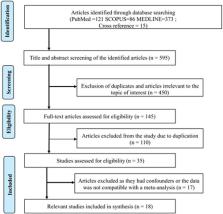
          The database search produced 595 articles from 2000 to 2022, among which 22 case reports were included in the systematic review. The mean age of JPOF occurrence in patients was 18 ± 16 years. A male predilection was noted among patients younger than 14 years of age, whereas a female predilection was noted in patients older than 14 years of age. Frequent involvement of the mandible (56%) compared to the maxilla (44%) was reported. The posterior mandible was the most commonly affected site involving numerous adjacent structures. The expansile nature of the JPOF displayed 57% buccolingual expansion, 50% downward displacement or erosion of the lower border of the mandible and 81% of  involvement of the maxillary antrum/pterygoid plate/orbital floor. Among the 20 cases reported, the treatment provided included surgical excision in 45% of the patients, jaw resection in 35% of the patients, and enucleation and curettage in 18% of the patients. Follow‐up details were provided in 80% of the reports that showed recurrence.

          Conclusions

          The diagnosis of JPOF requires correlation of the clinical and radiographic features with key histopathological features. Although long‐term follow‐up of the case reports has been reported, the data lack information about the long‐term outcomes of JPOF.

          Related collections

          Most cited references35

          • Record: found
          • Abstract: found
          • Article: not found

          Psammomatoid and trabecular juvenile ossifying fibroma of the craniofacial skeleton: two distinct clinicopathologic entities.

          The term juvenile ossifying fibroma is used in the literature in naming 2 microscopically distinct fibro-osseous lesions of the craniofacial skeleton. One is characterized by small uniform spherical ossicles resembling psammoma bodies (psammomatoid juvenile ossifying fibroma). The other is distinguished by trabeculae of fibrillary osteoid and woven bone (trabecular juvenile ossifying fibroma). Three new cases of each type are reported, and the literature is extensively reviewed for published cases of these 2 entities.Psammomatoid juvenile ossifying fibroma is reported more commonly than trabecular juvenile ossifying fibroma. It affects patients from a wider age range (3 months to 72 years) and an older mean age range (16-33 years) as compared with 2 to 12 years and 8(1/2) to 12 years, respectively, for trabecular juvenile ossifying fibroma. In both types there is a slight male predominance and the lesions are unencapsulated and tend to infiltrate adjacent bone. A significant difference between the 2 tumors is their site of occurrence. Although psammomatoid juvenile ossifying fibroma occurs predominantly in the sinonasal and orbital bones, trabecular juvenile ossifying fibroma predominantly affects the jaws. Aggressive growth occurs in some-but not all-cases of both types. Such behavior may be related to younger patient age and the concurrent development of aneurysmal bone cysts, which is seen more frequently in psammomatoid juvenile ossifying fibroma. This study demonstrates that not only histologic but also demographic and clinical differences between psammomatoid juvenile ossifying fibroma and trabecular juvenile ossifying fibroma warrant their classification as 2 distinct clinicopathologic entities.
            Bookmark
            • Record: found
            • Abstract: found
            • Article: not found

            Juvenile ossifying fibroma. An analysis of 33 cases with emphasis on histopathological aspects.

            Juvenile ossifying fibroma (JOF) is a maxillofacial fibro-osseous lesion that may show cell-rich osteoid strands or psammoma-like ossicles. Whether both types are variants of a single entity or different lesions under the same diagnostic label is a subject of debate. This problem was investigated by analyzing a series of 33 patients with lesions having one or both of the above histological appearances. It was concluded that osteoid strands define a unique fibro-osseous lesion but that lesions with psammoma-like ossicles fall within the morphological spectrum of cemento-ossifying fibroma. Therefore the term juvenile ossifying fibroma should be reserved for the lesion with the osteoid strands.
              Bookmark
              • Record: found
              • Abstract: not found
              • Book: not found

              Oral and Maxillofacial Pathology

                Bookmark

                Author and article information

                Contributors
                ghoshrocks2004@gmail.com
                Journal
                Clin Exp Dent Res
                Clin Exp Dent Res
                10.1002/(ISSN)2057-4347
                CRE2
                Clinical and Experimental Dental Research
                John Wiley and Sons Inc. (Hoboken )
                2057-4347
                03 November 2022
                February 2023
                : 9
                : 1 ( doiID: 10.1002/cre2.v9.1 )
                : 186-197
                Affiliations
                [ 1 ] Department of Oral Pathology Sathyabama Dental College and Hospital Chennai India
                [ 2 ] Department of Oral Pathology College of Medical Sciences Bharatpur Nepal
                [ 3 ] Department of Oral and Maxillofacial Surgery College of Medical Sciences Bharatpur Nepal
                Author notes
                [*] [* ] Correspondence Snehashish Ghosh, Department of Oral Pathology, College of Medical Sciences, Bharatpur, Nepal. 

                Email: ghoshrocks2004@ 123456gmail.com

                Author information
                http://orcid.org/0000-0002-7349-1619
                Article
                CRE2687
                10.1002/cre2.687
                9932254
                36325758
                4ef0e8f6-6714-47d4-8992-aa1ceb56cee8
                © 2022 The Authors. Clinical and Experimental Dental Research published by John Wiley & Sons Ltd.

                This is an open access article under the terms of the http://creativecommons.org/licenses/by/4.0/ License, which permits use, distribution and reproduction in any medium, provided the original work is properly cited.

                History
                : 20 October 2022
                : 27 September 2022
                : 22 October 2022
                Page count
                Figures: 1, Tables: 2, Pages: 12, Words: 5713
                Categories
                Review Article
                Review Article
                Custom metadata
                2.0
                February 2023
                Converter:WILEY_ML3GV2_TO_JATSPMC version:6.2.5 mode:remove_FC converted:16.02.2023

                fibroma,juvenile,ossifying,psammomatoid
                fibroma, juvenile, ossifying, psammomatoid

                Comments

                Comment on this article