37
views
0
recommends
+1 Recommend
0 collections
    0
    shares
      • Record: found
      • Abstract: found
      • Article: found
      Is Open Access

      LMP1-mediated glycolysis induces myeloid-derived suppressor cell expansion in nasopharyngeal carcinoma

      research-article

      Read this article at

      Bookmark
          There is no author summary for this article yet. Authors can add summaries to their articles on ScienceOpen to make them more accessible to a non-specialist audience.

          Abstract

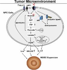
          Myeloid-derived suppressor cells (MDSCs) are expanded in tumor microenvironments, including that of Epstein–Barr virus (EBV)-associated nasopharyngeal carcinoma (NPC). The link between MDSC expansion and EBV infection in NPC is unclear. Here, we show that EBV latent membrane protein 1 (LMP1) promotes MDSC expansion in the tumor microenvironment by promoting extra-mitochondrial glycolysis in malignant cells, which is a scenario for immune escape initially suggested by the frequent, concomitant detection of abundant LMP1, glucose transporter 1 (GLUT1) and CD33 + MDSCs in tumor sections. The full process has been reconstituted in vitro. LMP1 promotes the expression of multiple glycolytic genes, including GLUT1. This metabolic reprogramming results in increased expression of the Nod-like receptor family protein 3 (NLRP3) inflammasome, COX-2 and P-p65 and, consequently, increased production of IL-1β, IL-6 and GM-CSF. Finally, these changes in the environment of malignant cells result in enhanced NPC-derived MDSC induction. One key step is the physical interaction of LMP1 with GLUT1 to stabilize the GLUT1 protein by blocking its K48-ubiquitination and p62-dependent autolysosomal degradation. This work indicates that LMP1-mediated glycolysis regulates IL-1β, IL-6 and GM-CSF production through the NLRP3 inflammasome, COX-2 and P-p65 signaling pathways to enhance tumor-associated MDSC expansion, which leads to tumor immunosuppression in NPC.

          Author summary

          The expression of the Epstein-Barr virus (EBV) oncogenic protein denoted latent membrane protein 1 (LMP1) varies in patients with NPC and is linked to tumorigenesis and tumor immunosuppression, but the molecular mechanism through which LMP1 leads to tumor immune escape remains unknown. Work to date suggests that the expansion of tumor-associated myeloid-derived suppressor cells (MDSCs) is the main cause of tumor immunosuppression such as that found in NPC. Here, we found that tumor LMP1 expression is correlated with glucose transporter 1 (GLUT1) levels, CD33 + MDSC number and unfavorable survival in patients with NPC. Based on the results of our in vitro analysis, LMP1 promotes GLUT1-dependent glycolysis in NPC cells, resulting in activation of the Nod-like receptor family protein 3 (NLRP3) inflammasome, COX-2 and P-p65 signaling pathways and subsequently increased IL-1β, IL-6 and GM-CSF production. Importantly, LMP1 interacts with GLUT1 to stabilize the GLUT1 protein by disrupting its K48-linked ubiquitination and autolysosomal degradation in a p62-dependent manner and up-regulating the GLUT1 mRNA and protein levels by inducing p65 activation. Therefore, we determined that GLUT1-dependent glycolysis is required for tumor-induced MDSC differentiation and that this process is associated with LMP1 expression. Based on our findings, LMP1-mediated glycolysis is a key process involved in controlling tumor immunosuppression and directly contributes to oncogenesis.

          Related collections

          Most cited references48

          • Record: found
          • Abstract: found
          • Article: not found

          Modulation of microenvironment acidity reverses anergy in human and murine tumor-infiltrating T lymphocytes.

          Stimulating the effector functions of tumor-infiltrating T lymphocytes (TIL) in primary and metastatic tumors could improve active and adoptive T-cell therapies for cancer. Abnormal glycolysis, high lactic acid production, proton accumulation, and a reversed intra-extracellular pH gradient are thought to help render tumor microenvironments hostile to roving immune cells. However, there is little knowledge about how acidic microenvironments affect T-cell immunity. Here, we report that lowering the environmental pH to values that characterize tumor masses (pH 6-6.5) was sufficient to establish an anergic state in human and mouse tumor-specific CD8(+) T lymphocytes. This state was characterized by impairment of cytolytic activity and cytokine secretion, reduced expression of IL-2Rα (CD25) and T-cell receptors (TCR), and diminished activation of STAT5 and extracellular signal-regulated kinase (ERK) after TCR activation. In contrast, buffering pH at physiologic values completely restored all these metrics of T-cell function. Systemic treatment of B16-OVA-bearing mice with proton pump inhibitors (PPI) significantly increased the therapeutic efficacy of both active and adoptive immunotherapy. Our findings show that acidification of the tumor microenvironment acts as mechanism of immune escape. Furthermore, they illustrate the potential of PPIs to safely correct T-cell dysfunction and improve the efficacy of T-cell-based cancer treatments. ©2012 AACR
            Bookmark
            • Record: found
            • Abstract: found
            • Article: not found

            Characterization of cytokine-induced myeloid-derived suppressor cells from normal human peripheral blood mononuclear cells.

            Tumor immune tolerance can derive from the recruitment of suppressor cell populations, including myeloid-derived suppressor cells (MDSCs). In cancer patients, increased MDSCs correlate with more aggressive disease and a poor prognosis. Expression of 15 immune factors (TGFbeta, IL-1beta, IL-4, IL-6, IL-10, GM-CSF, M-CSF, IDO, fms-related tyrosine kinase 3 ligand, c-kit ligand, inducible NO synthase, arginase-1, TNF-alpha, cyclo-oxygenase 2, vascular endothelial growth factor [VEGF]) by MDSC-inducing human solid tumor cell lines was evaluated by RT-PCR. Based upon these data, cytokine mixtures were then tested for their ability to generate suppressive CD33(+) cells from healthy donor PBMCs in vitro by measuring their ability to inhibit the proliferation of, and IFN-gamma production by, fresh autologous human T cells after CD3/CD28 stimulation. Induced MDSCs were characterized with respect to their morphology, surface phenotype, and gene expression profile. MDSC-inducing cancer cell lines demonstrated multiple pathways for MDSC generation, including overexpression of IL-6, IL-1beta, cyclo-oxygenase 2, M-CSF, and IDO. CD33(+) cells with potent suppressive capacity were best generated in vitro by GM-CSF and IL-6, and secondarily by GM-CSF + IL-1beta, PGE(2), TNF-alpha, or VEGF. Characterization studies of cytokine-induced suppressive cells revealed CD33(+)CD11b(+)CD66b(+)HLA-DR(low)IL-13R alpha2(int) large mononuclear cells with abundant basophilic cytoplasm. Expression of inducible NO synthase, TGFbeta, NADPH oxidase, VEGF, and/or arginase-1 was also upregulated, and Transwell studies showed suppression of autologous T cells to be contact dependent. Suppressive CD33(+) cells generated from PBMCs by GM-CSF and IL-6 were consistent with human MDSCs. This study suggests that these cytokines are potential therapeutic targets for the inhibition of MDSC induction in cancer patients.
              Bookmark
              • Record: found
              • Abstract: found
              • Article: found
              Is Open Access

              PKM2-dependent glycolysis promotes NLRP3 and AIM2 inflammasome activation

              Sepsis, severe sepsis and septic shock are the main cause of mortality in non-cardiac intensive care units. Immunometabolism has been linked to sepsis; however, the precise mechanism by which metabolic reprogramming regulates the inflammatory response is unclear. Here we show that aerobic glycolysis contributes to sepsis by modulating inflammasome activation in macrophages. PKM2-mediated glycolysis promotes inflammasome activation by modulating EIF2AK2 phosphorylation in macrophages. Pharmacological and genetic inhibition of PKM2 or EIF2AK2 attenuates NLRP3 and AIM2 inflammasomes activation, and consequently suppresses the release of IL-1β, IL-18 and HMGB1 by macrophages. Pharmacological inhibition of the PKM2–EIF2AK2 pathway protects mice from lethal endotoxemia and polymicrobial sepsis. Moreover, conditional knockout of PKM2 in myeloid cells protects mice from septic death induced by NLRP3 and AIM2 inflammasome activation. These findings define an important role of PKM2 in immunometabolism and guide future development of therapeutic strategies to treat sepsis.
                Bookmark

                Author and article information

                Contributors
                Role: Data curationRole: MethodologyRole: Writing – original draftRole: Writing – review & editing
                Role: Data curationRole: Formal analysisRole: Methodology
                Role: Data curationRole: Methodology
                Role: Data curationRole: Methodology
                Role: Data curationRole: Resources
                Role: Data curationRole: Resources
                Role: Data curationRole: Methodology
                Role: Data curationRole: ResourcesRole: Writing – review & editing
                Role: Resources
                Role: VisualizationRole: Writing – review & editing
                Role: Resources
                Role: ConceptualizationRole: Data curationRole: Formal analysisRole: Funding acquisitionRole: InvestigationRole: MethodologyRole: ResourcesRole: SupervisionRole: VisualizationRole: Writing – original draftRole: Writing – review & editing
                Role: Editor
                Journal
                PLoS Pathog
                PLoS Pathog
                plos
                plospath
                PLoS Pathogens
                Public Library of Science (San Francisco, CA USA )
                1553-7366
                1553-7374
                21 July 2017
                July 2017
                : 13
                : 7
                : e1006503
                Affiliations
                [1 ] Collaborative Innovation Center for Cancer Medicine, State Key Laboratory of Oncology in South China, Guangzhou, China
                [2 ] Guangdong Provincial Key Laboratory of Colorectal and Pelvic Floor Diseases, The Sixth Affiliated Hospital, Sun Yat-sen University, Guangzhou, China
                [3 ] Department of Biotherapy, Sun Yat-sen University Cancer Center, Guangzhou, China
                [4 ] Department of Nasopharyngeal Carcinoma, Sun Yat-sen University Cancer Center, Guangzhou, China
                [5 ] Key Laboratory of Gene Engineering of the Ministry of Education, State Key Laboratory of Biocontrol, College of Life Sciences, Sun Yat-sen University, Guangzhou, China
                [6 ] Université Paris-Sud-11, CNRS-UMR 8126 and Institut de Cancérologie Gustave Roussy, 39 rue Camille Desmoulins, Villejuif, France
                Northwestern University, UNITED STATES
                Author notes

                The authors have declared that no competing interests exist.

                Author information
                http://orcid.org/0000-0003-1027-3400
                http://orcid.org/0000-0002-7243-4671
                Article
                PPATHOGENS-D-17-00235
                10.1371/journal.ppat.1006503
                5540616
                28732079
                4bd9dac8-8013-4182-84e2-7ca493b5fa28
                © 2017 Cai et al

                This is an open access article distributed under the terms of the Creative Commons Attribution License, which permits unrestricted use, distribution, and reproduction in any medium, provided the original author and source are credited.

                History
                : 3 February 2017
                : 30 June 2017
                Page count
                Figures: 7, Tables: 0, Pages: 23
                Funding
                Funded by: funder-id http://dx.doi.org/10.13039/501100001809, National Natural Science Foundation of China;
                Award ID: 2013CB910301
                Award Recipient :
                Funded by: funder-id http://dx.doi.org/10.13039/501100001809, National Natural Science Foundation of China;
                Award ID: 81572982, 81372442 and 81172164
                Award Recipient :
                Funded by: Guangdong Province Science Foundation
                Award ID: 2014A020212066
                Award Recipient :
                This work was supported by the National Basic Research Program of China (2013CB910301, YZ), the General Program (Grant Nos. 81572982, 81372442 and 81172164, JL) of the National Natural Science Foundation of China ( http://www.nsfc.gov.cn/), and the Sci-Tech Key Program of the Guangdong Province Science Foundation (Grant No. 2014A020212066, JL) http://pro.gdstc.gov.cn/egrantweb/. The funders had no role in study design, data collection and analysis, decision to publish, or preparation of the manuscript.
                Categories
                Research Article
                Biology and Life Sciences
                Biochemistry
                Metabolism
                Metabolic Processes
                Glycolysis
                Research and Analysis Methods
                Precipitation Techniques
                Immunoprecipitation
                Biology and Life Sciences
                Immunology
                Immune System Proteins
                Inflammasomes
                Medicine and Health Sciences
                Immunology
                Immune System Proteins
                Inflammasomes
                Biology and Life Sciences
                Biochemistry
                Proteins
                Immune System Proteins
                Inflammasomes
                Research and Analysis Methods
                Immunologic Techniques
                Immunoassays
                Enzyme-Linked Immunoassays
                Biology and Life Sciences
                Biochemistry
                Metabolism
                Carbohydrate Metabolism
                Glucose Metabolism
                Medicine and Health Sciences
                Oncology
                Cancers and Neoplasms
                Carcinomas
                Nasopharyngeal Carcinoma
                Physical Sciences
                Chemistry
                Chemical Compounds
                Organic Compounds
                Carbohydrates
                Monosaccharides
                Glucose
                Physical Sciences
                Chemistry
                Organic Chemistry
                Organic Compounds
                Carbohydrates
                Monosaccharides
                Glucose
                Biology and Life Sciences
                Physiology
                Immune Physiology
                Cytokines
                Medicine and Health Sciences
                Physiology
                Immune Physiology
                Cytokines
                Biology and Life Sciences
                Immunology
                Immune System
                Innate Immune System
                Cytokines
                Medicine and Health Sciences
                Immunology
                Immune System
                Innate Immune System
                Cytokines
                Biology and Life Sciences
                Developmental Biology
                Molecular Development
                Cytokines
                Custom metadata
                vor-update-to-uncorrected-proof
                2017-08-02
                All relevant data are within the paper and its Supporting Information files.

                Infectious disease & Microbiology
                Infectious disease & Microbiology

                Comments

                Comment on this article