5
views
0
recommends
+1 Recommend
0 collections
    0
    shares
      • Record: found
      • Abstract: found
      • Article: found
      Is Open Access

      Oxeiptosis: a novel pathway of melanocytes death in response to oxidative stress in vitiligo

      research-article

      Read this article at

      Bookmark
          There is no author summary for this article yet. Authors can add summaries to their articles on ScienceOpen to make them more accessible to a non-specialist audience.

          Abstract

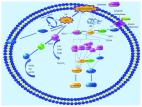
          Vitiligo is a cutaneous depigmenting autoimmune disease caused by the extensive destruction of epidermal melanocytes. Convincing data has defined a critical role for oxidative stress in the pathogenesis of vitiligo. Oxeiptosis is a caspase-independent cell death modality that was reportedly triggered by oxidative stress and operative in pathogen clearance. However, whether oxeiptosis exists in oxidative stress-induced melanocytes demise in vitiligo remains undetermined. In the present study, we initially found that other cell death modalities might exist in addition to the well-recognized apoptosis and necroptosis in H 2O 2-treated melanocytes. Furthermore, AIFM1 was found to be dephosphorylated at Ser116 in oxidative stress-induced melanocytes death, which was specific to oxeiptosis. Moreover, KEAP1 and PGAM5, upstream of the AIFM1 in oxeiptosis, were found to operate in melanocytic death. Subsequently, the KEAP1-PGAM5-AIFM1 signaling pathway was proved to be involved in oxidative stress-triggered melanocytes demise through the depletion of KEAP1 and PGAM5. Altogether, our study indicated that oxeiptosis might occur in melanocytes death under oxidative stress and contribute to the pathogenesis of vitiligo.

          Related collections

          Most cited references38

          • Record: found
          • Abstract: found
          • Article: found
          Is Open Access

          Reactive Oxygen Species-Induced Lipid Peroxidation in Apoptosis, Autophagy, and Ferroptosis

          Reactive oxygen species- (ROS-) induced lipid peroxidation plays a critical role in cell death including apoptosis, autophagy, and ferroptosis. This fundamental and conserved mechanism is based on an excess of ROS which attacks biomembranes, propagates lipid peroxidation chain reactions, and subsequently induces different types of cell death. A highly evolved sophisticated antioxidant system exists that acts to protect the cells from oxidative damage. In this review, we discussed how ROS propagate lipid peroxidation chain reactions and how the products of lipid peroxidation initiate apoptosis and autophagy in current models. We also discussed the mechanism of lipid peroxidation during ferroptosis, and we summarized lipid peroxidation in pathological conditions of critical illness. We aim to bring a more global and integrative sight to know how different ROS-induced lipid peroxidation occurs among apoptosis, autophagy, and ferroptosis.
            Bookmark
            • Record: found
            • Abstract: found
            • Article: found
            Is Open Access

            ROS and the DNA damage response in cancer

            Reactive oxygen species (ROS) are a group of short-lived, highly reactive, oxygen-containing molecules that can induce DNA damage and affect the DNA damage response (DDR). There is unequivocal pre-clinical and clinical evidence that ROS influence the genotoxic stress caused by chemotherapeutics agents and ionizing radiation. Recent studies have provided mechanistic insight into how ROS can also influence the cellular response to DNA damage caused by genotoxic therapy, especially in the context of Double Strand Breaks (DSBs). This has led to the clinical evaluation of agents modulating ROS in combination with genotoxic therapy for cancer, with mixed success so far. These studies point to context dependent outcomes with ROS modulator combinations with Chemotherapy and radiotherapy, indicating a need for additional pre-clinical research in the field. In this review, we discuss the current knowledge on the effect of ROS in the DNA damage response, and its clinical relevance.
              Bookmark
              • Record: found
              • Abstract: found
              • Article: not found

              The KEAP1-NRF2 System: a Thiol-Based Sensor-Effector Apparatus for Maintaining Redox Homeostasis.

              The Kelch-like ECH-associated protein 1-NF-E2-related factor 2 (KEAP1-NRF2) system forms the major node of cellular and organismal defense against oxidative and electrophilic stresses of both exogenous and endogenous origins. KEAP1 acts as a cysteine thiol-rich sensor of redox insults, whereas NRF2 is a transcription factor that robustly transduces chemical signals to regulate a battery of cytoprotective genes. KEAP1 represses NRF2 activity under quiescent conditions, whereas NRF2 is liberated from KEAP1-mediated repression on exposure to stresses. The rapid inducibility of a response based on a derepression mechanism is an important feature of the KEAP1-NRF2 system. Recent studies have unveiled the complexities of the functional contributions of the KEAP1-NRF2 system and defined its broader involvement in biological processes, including cell proliferation and differentiation, as well as cytoprotection. In this review, we describe historical milestones in the initial characterization of the KEAP1-NRF2 system and provide a comprehensive overview of the molecular mechanisms governing the functions of KEAP1 and NRF2, as well as their roles in physiology and pathology. We also refer to the clinical significance of the KEAP1-NRF2 system as an important prophylactic and therapeutic target for various diseases, particularly aging-related disorders. We believe that controlled harnessing of the KEAP1-NRF2 system is a key to healthy aging and well-being in humans.
                Bookmark

                Author and article information

                Contributors
                lishli@fmmu.edu.cn
                Journal
                Cell Death Discov
                Cell Death Discov
                Cell Death Discovery
                Nature Publishing Group UK (London )
                2058-7716
                17 February 2022
                17 February 2022
                2022
                : 8
                : 70
                Affiliations
                GRID grid.233520.5, ISNI 0000 0004 1761 4404, Department of Dermatology, Xijing Hospital, , Fourth Military Medical University, ; No. 127 of West Changle Road, Xi’an, Shaanxi 710032 China
                Author information
                http://orcid.org/0000-0003-3004-5376
                http://orcid.org/0000-0002-2335-507X
                Article
                863
                10.1038/s41420-022-00863-3
                8854565
                35177586
                4b2f928b-692a-4891-8288-be844280f54d
                © The Author(s) 2022

                Open Access This article is licensed under a Creative Commons Attribution 4.0 International License, which permits use, sharing, adaptation, distribution and reproduction in any medium or format, as long as you give appropriate credit to the original author(s) and the source, provide a link to the Creative Commons license, and indicate if changes were made. The images or other third party material in this article are included in the article’s Creative Commons license, unless indicated otherwise in a credit line to the material. If material is not included in the article’s Creative Commons license and your intended use is not permitted by statutory regulation or exceeds the permitted use, you will need to obtain permission directly from the copyright holder. To view a copy of this license, visit http://creativecommons.org/licenses/by/4.0/.

                History
                : 3 November 2021
                : 18 January 2022
                : 1 February 2022
                Funding
                Funded by: FundRef https://doi.org/10.13039/501100001809, National Natural Science Foundation of China (National Science Foundation of China);
                Award ID: 81803122
                Award ID: 81803141
                Award ID: 81930087
                Award ID: 91742201
                Award ID: 81625020
                Award Recipient :
                Categories
                Article
                Custom metadata
                © The Author(s) 2022

                chronic inflammation,cell death
                chronic inflammation, cell death

                Comments

                Comment on this article